Poster
# 53
![]()
Poster |
|
6th Internet World Congress for Biomedical Sciences |
Eiichi Yagi(1)
(1)Akita Red Cross Hospital - Akita. Japan
|
|
|
|
|
|
[Dermatology] |
[Health Informatics] |
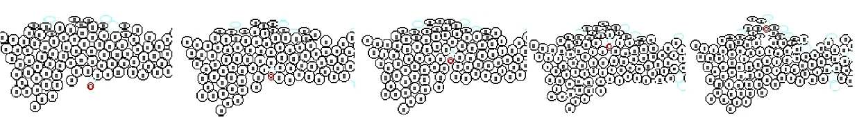
The virtual epidermal cells on the screen (in the computer memory) were multiplied with changing the various kinds of parameter in these cells, and the morphological change of the total structure after growth was examined.
Hardware and soft ware: PC-AT compatible computer AMD K2-450 (RAM 256MB)
video RAM 16M, Windows 98,
Visual Basic 5.0 (Microsoft)
( All programs were written by Eiichi Yagi MD) Virtual cells multiply as a kind of celler automaton algorithm. (Fig. 1)
1- distance of all cells from initial the dividing cell is measured.
2- new cell keeps 1dot from the dividing cell.
3- extract the nearest cell that overlapped with the dividing cell.
4- these cells are supposed to be a ball, then the direction of the move is calculated.
5- the position of cells is corrected not to be overlapped.
6- the cells that overlapped with this cell are computed, and process 4 is done with all cells that are extracted.
7- return to process 2, and the work has been done until all cells are completely divided.
The form of virtual epidermis changes with regulating the selection probability, the mitosis probability, the radius and division direction.
Figure 1
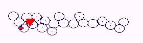
Celler automaton
This is an automatic proliferation model devised by Neumann. Unit automaton that is arranged on the plane like a cell changes its internal state only according to the condition of self and its vicinity, so the movement of all units automatically determines the movement of the whole system.
Setting of the parameter and the function of the individual virtual cell
X-coordinate, Y-coordinate, radius, a longer axis, a division tendency, display of division direction, on basal layer or not, Xb of cell (growth factor), Yb of cell (inhibitory growth factor), a history of the successive division which depended on concentration, initial width, initial height, cell number, basal layer - x,y, granular layer - x,y, distance to the horny layer, division frequency, a size difference of a cell in division, division angle after division, division frequency with the same range, division arrest after successive division, elasticity on the horny layer, elimination frequency on the horny layer, elimination frequency on theh horny layer (altitude is not related), How many horny layers is torn off?, only basal cell is divided, probability that high concentration induced the division on the basal layer, ratio of diffusion rate of Xb and Yb, increse of Xb substance synthesis, decrease of Xb substance synthesis, division direction on the basal layer - level, verticality, angle
Random control for each parameter -- another 10-15 parameters are required.
Functions for the internal processing in proliferation -- another 10-15 functions are required.
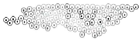
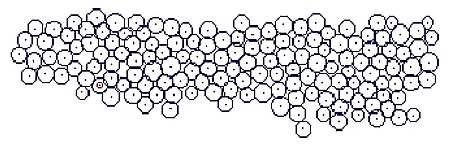
Simple growth
Figure 2
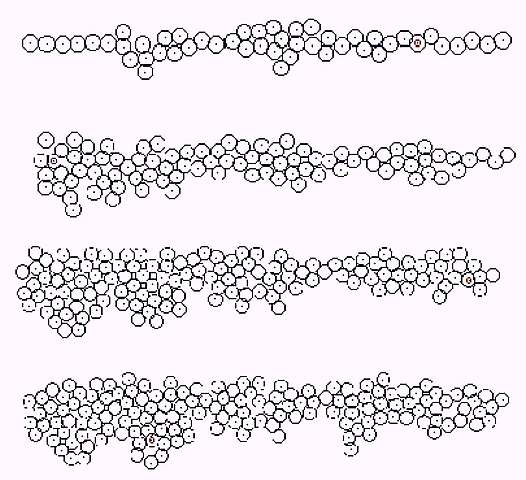
When the growth rate of surroundings cells selected for growth was increased, the rete ridge like structure of was made (Fig. 2). Rete ridge seemed to be longer when the division probability of the basal layer increased.
Applied case of growth
P (growth factor) and Q (Inhibition factor) were put in cells according to Turing method. Concentration (P) was displayed by the darkness of cell when R (diffusion rate) was regulated appropriately. The cells, which were with high concentration and near to basal layer, were divided with precedence.
Figure 3
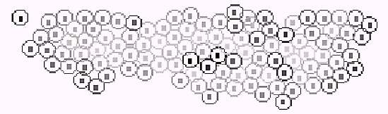
By setting of coefficients, P (growth factor) was arranged in layered formation. Virtual epidermis tends to thicken according to the increase of P (Fig 3). P was able to move toward outside by another coefficient setting. Dilatation speed of eruption was controlled with the balance of P, Q and R.
Figure 4
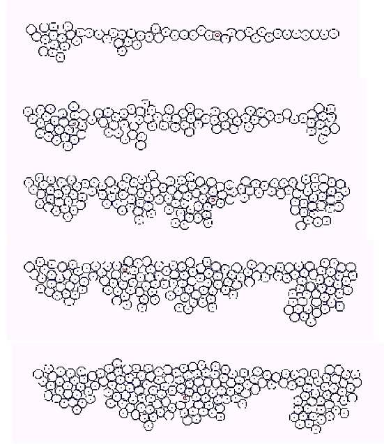
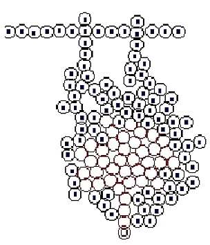
Cases of proliferation
The switch controlling the proliferation in the lower part of basal layer was set by P, Q factor and the division frequency. Then the structure as a hair root was made (Fig. 5).
Figure 5
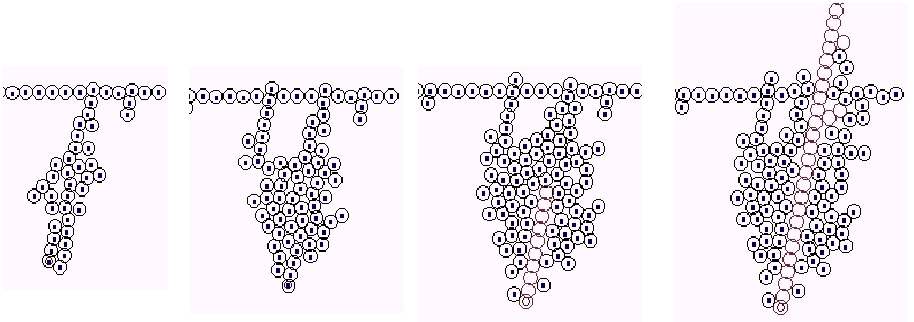
A difference of cell size was set large, and a division position was set over the basal layer. It showed like atypicality in situ (in the epidermis)(Fig. 6). Fractal dimension of circumference borderline increases as the probability of difference of cell size rises.
Figure 6
When the division directionality and the adherence to circumference of virtual cell are regulated, a cyst-like structure is constructed (Fig. 7). Keratinization can be done from upward of the tumor. When the direction or speed of the initial keratinization is in a certain range, the structure may be destructed and absorbed immediately.
Figure 7
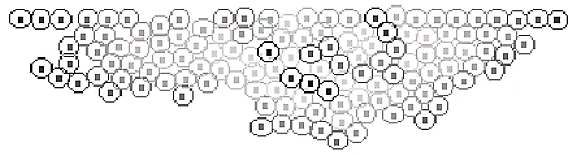
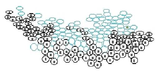
Cases of proliferation -- addition of horny and granular layer
Virtual granular layer (differentiation stage) is added (Fig. 8), and keratinization is occurred in random position. Virtual granule cells are shown as oval cells, and horny cells are shown as light blued oval cells.
Figure 8

Increase of keratinization tendency and difference of size are added (Fig. 9).
Figure 9
Addition of the randomized differentiation phase and the difference of size (Fig. 10)
Figure 10
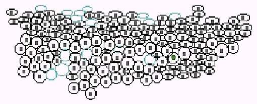
Addition of virtual infiltrate cell (Fig. 11)
Similar structure as subcorneal abscess can be made by this option. Implantation of acceleration / inhibiting factor of proliferation in this cell is possible.
Figure 11
By using this growth model the relationship between the arrangement of cellular level and the mass/fractal dimension of structure and the configuration dimension of macro-construction can be examined. If z-axis is added to the functions, the extension to three-dimensional structure is possible. However, the computation time may become enormous.
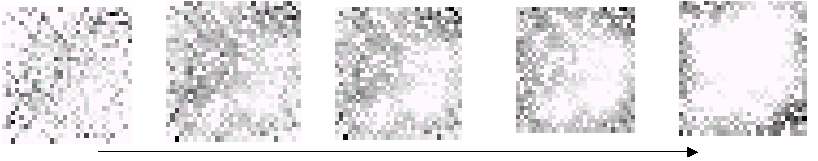
Image processing of histopathological data
Figure 12

These images were automatically made, and the dimensions were
simultaneously calculated by the system ( View1 1.1 made by E Yagi)
Making of time series data (making of 700-2000 vectors )
1)Distance between cells
a. Cell interval is sought when the marker is arranged in concentric circular from the center of the image.
b. All combinations of distance between all 2 cells are sought, and the cell number that two cells enter in the limited interval divided with equal length is counted.
c. Cells are plotted with a certain width lengthwise, and arranged in order from near side. Distance of all pairs is measured.
2)Size of nucleus, A length / area of circumference, Concentration difference of nucleus
Value is put away in order of coordinate, and nucleus is extracted by order of 1).
Specimens to examine histopathologically
normal skin, seborrheic keratosis 6 cases, actinic keratosis 8 cases, Bowen´s disease 6 cases, basal cell carcinoma 5 cases, squamous cell carcinoma 4 cases, Paget´s disease 4 cases, dermatofibroma 3 cases, hemangioma 4 cases, pigmented nevus 6 cases
| Distance between cells | Size of nucleus | A length / area of circumference | Concentration difference of nucleus | |
| normal skin | 6.1 | 3.31 | 6.0 | 5.92 |
| seborrheic keratosis | 5.1 | 4.3 | 5.82 | 5.34 |
| actinic keratosis | 5.0 | 2.61 | 6.4 | 4.2 |
| Bowen disease | 5.0 | 1.42 | 6.75 | 4.02 |
| squamous cell carcinoma | 4.9 | 0.85 | 7.87 | 6.15 |
| basal cell cancer | 5.2 | 1.35 | 6.67 | 1.74 |
| Paget disease | 4.6 | 2.65 | 7.4 | 3.1 |
| dermatofibroma | 4.84 | 2.6 | 7.68 | 6.88 |
| hemangioma | 3.6 | 2.22 | 6.8 | 7.9 |
| pigmented nevus | 6.05 | 3.0 | 6.16 | 6.2 |
| Simple fractal dimension of the circumference in virtual tumor |  |
| Probability of the cell size change at one division of cell (0 to 0.9) |
In order to bring the character of virtual tumor close to the real tumor the coincidence of these dimensions is necessary. Coincidence within a small range may be possible, although a lot of computation process is necessary even in such accordance. If various kinds of algorithms (Genetic Algorithms, serious inquiry method etc.) are used and applied appropriately to choose the functions and coefficients, the examination of character with spreading of cells may be possible.
The unique characteristics of the total structure were given by changing the parameters and the functions of virtual cell, and the virtual eruption derived from dilatation tendency and the various kinds of form were made by the delicate and ideal balance control of parameters. This system of making virtual skin tumors may be useful as the apparatus, which investigates characteristic and relationship between macro and microstructure.
|
|
|
|
|
|
[Dermatology] |
[Health Informatics] |