Póster
Nş 065

Póster |
|
Giorgio Gherardi, M.D., Andrea Baiocchini, M.D., Piero Di Zitti, M.D.
![]()
|
|
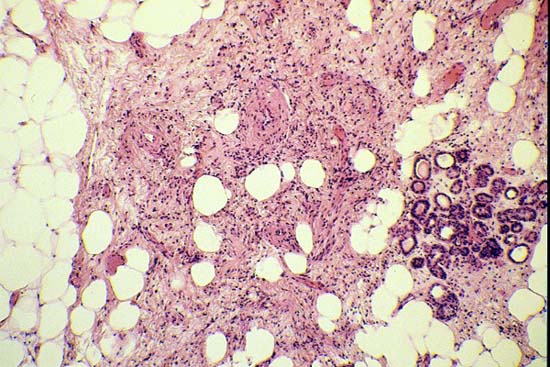 |
| Fig. 1 : The lesion consisted of newly formed abnormal blood vessels, spindly smooth muscle cells accentuating around vessels, and mature adipocytes. Note that in this field the proliferation is in close contact with a mammary lobule. Haematoxylin-eosin, x150. |
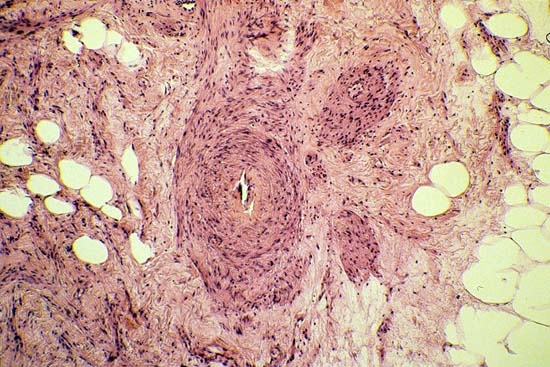 |
| Fig. 2 : Blood vessels frequently had the shape of small arteries and showed subintimal fibrosis ; smooth muscle cells surrounded such vessels and appeared to depart from them. Haematoxylin-eosin, x150 |
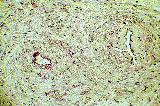 |
| Fig. 3 : Two vessels are illustrated after histochemical Weigert staining for elastica. While one vessel is characterized by an elastic lamina, the other has no elastica and displays an irregularly shaped lumen. Haemotoxylin-eosin, x200 |
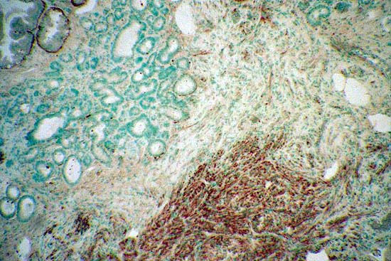 |
| Fig. 4 : Spindle cells show positive immunoreactivity for smooth muscle actin. Adjacent mammary glands also show positive staining. ABC immunoperoxidase technique, x180 |
|
|