Póster
Nş 065

Póster |
|
Giorgio Gherardi, M.D., Andrea Baiocchini, M.D., Piero Di Zitti, M.D.
![]()
|
|
The neoplasm we described here shows pathologic features that fulfill the diagnostic criteria of AML ( 4 ). In fact it was characterized by the mixed proliferation of three components represented by smooth muscle, blood vessels, and mature fat. As in AML the smooth muscle cells were arranged in fascicles and bundles that accentuated around blood vessels. These were characteristically thick walled, tortuous, with little or no elastic lamina and showed subintimal fibrosis, and, finally, fat cells had the morphologic features of mature adipocytes.
In our opinion only angiolipoma ( 5 ) and myoid hamarthoma ( 6 ) of the breast enter the differential diagnosis of the present lesion. Since, however, the currently reported neoplasm was characterized by a mixed proliferation of the above three cellular components, both entities can be excluded due to the lack of a muscular component in angiolipoma ( 5 ) and of a vascular component in myoid hamarthoma ( 6 ). Finally, the characteristic morphology of abnormal vessels in the present case strongly favours the diagnosis of AML ( 4 ).
Whether AML represents a hamarthomatous or a neoplastic process is still a matter of debate. According to Bonetti et al ( 1 ) the tumor would originate from a single cell type with pluripotential ability, probably to be identified as the so-called “perivascular epithelioid cell”. This latter is characterized by the immunohistochemical coexpression of muscle (actin and, less frequently, desmin) and melanogenesis markers (HMB-45 ). In AML melanogenesis markers are expressed by a peculiar component of bizarre and epithelioid cells which are commonly well developed in renal or hepatic tumors but are usually absent in AML arising in other locations ( 6-10 ). It is for this reason that the diagnosis of AML in extrarenal and extrahepatic sites should rely on the peculiar findings of a mixed proliferation of the above components and on the characteristic abnormalities of blood vessels which are unique to this tumor while immunohistochemical search of melanogenesis markers commonly gives negative results ( 6-10 ).
Renal AML can also present as a multicentric tumor ( 1,4 ). To rule out the chance that the mammary lesion could represent a distant location of a renal AML with multicentric lesions we reviewed the pathologic specimens of the left kidney previously removed for treatment of a renal cell carcinoma to rule out the chance of a coexistent AML possibly overlooked at the time of this latter diagnosis, and advised that the patient underwent a CT-scan of the contralateral kidney. Both investigations gave negative results thus demonstrating that the tumor represented a primary and totally isolated form of AML in the breast.
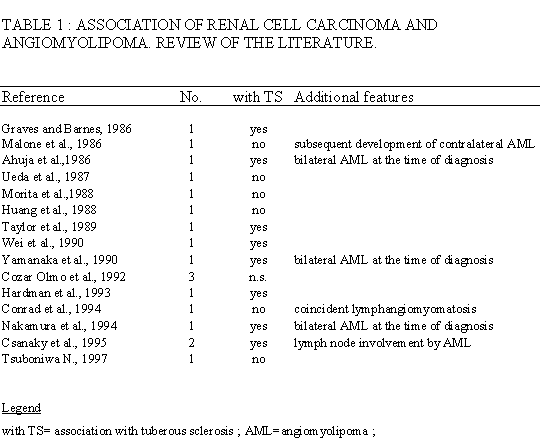
Primary extrarenal AMLs are very rare and no more than 50 cases have been reported in the literature. Extrarenal locations include hepatobiliary and reproductive tracts, lung, heart, nasal cavity, hard palate, abdominal wall, skin, colon, and retroperitoneum ((for review see Fegan et al. (2)). The case under study would show an additional peculiarity which is represented by the development of AML in a patient recently treated for clear cell renal carcinoma. The association of AML and renal cell carcinoma is a very rare but well known phenomenon since at least 18 cases have been reported in the literature (see Table 1). The association cannot be coincidental since both AML and renal cell carcinoma are strongly associated with tuberous sclerosis and, in fact, in nine out of the cases listed in Table 1 the two neoplasms were detected in this setting. The present case can be added to the above series and bears the peculiarity of being the first reported case of AML in a patient with renal cell carcinoma that developed in an extrarenal location.
|
|