Poster
# 104
![]()
Poster |
|
6th Internet World Congress for Biomedical Sciences |
Klimentina Nesterova(1), Ivan Nesterov(2)
(1)Omsk state medical Academy - Omsk. Russian Federation
(2)Omsk Medical Academy - Omsk. Russian Federation
|
|
|
|
|
|
[Infectious Diseases] |
[Otorhinolaryngology] |
It´s common knowledge, that clinical current of acute and chronic diseases of upper respiratory tract is mostly stipulated by the characteristics of the microbial flora of the nasal cavity and the amount of stamms, resistant to the medicines, used for treatment. The microbe landscape of nasal mucous has considerable age and regional particularities.
We´ve investigated the microbe flora of nasal exudations on the different stages of bacterial rhinitis beside 659 patients and determined it´s cultural characteristics, taxonomic position, microbal number and sensitivity to different antibiotics and some antiseptics. Amongst the patients there are 399 adults and 280 children under 14 years old. During our investigation 679 bacteriological analyses were done and 678 of them showed the presence of microorganisms (in 1 (0.15%) sowing there were no bacterial flora in the analyzed material). In 20 cases microbes were found in associations, the most often was the association of staphylococcus with funguses. In 3 patients we´ve found an association of three microorganisms, in one case- 4 microorganisms: Pseudomonas aerugenosa + Candida crusei + Aspergillus niger + Aspergillus flava. The most often microorganism was Staphylococcus. We´ve found it in 72 % of all sowings. In 18 cases (3 % of all sowings where staphylococcus were found) it was in association with other microorganisms.
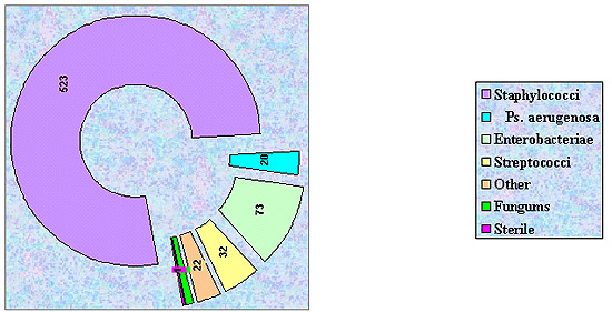
Diagram 1. Aspectual distribution of microflora of nasal cavities.
More then a half of the group was represented by Staphylococcus epidermidis, it was found in 40,64% of all samples, received from nasal cavity. Besides, it was found in adults 1,5 times more often (61.5%) then in children (40,7%).
In age group "under 14 years» we´ve noticed the domination of Staphylococcus aureus in nasal exudation, it was found 2 times more often in children, then in adults. We decided, that exactly S. aureus, found in 40 % of all children wit bacterial rhinitis, is the main perpetrator of stubborn relapsing of this disease in this age, as more aggressive then S. epidermidis or S. saprophyticus.
The last one represented only 10 % of all staphylococci and is evenly distributed between adults and children.
Table 1
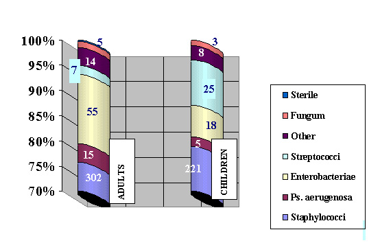
Diagram 2. Aspectual distribution of nasal cavity microflora under the age aspect.
Enterobacteriums are represented in vastly smaller amount. They were found only in 73 analyses (10,8 %). Every species was found not more then in 1,5- 2 % of all patients. Mostly they are found in adults (2 times more often), but Proteus vulgaris, Esherichia coli and Clebsiella pneumoniae are equally distributed between the age groups. In adults we found a wide variety of Proteae tribe: Pr. mirabilis 7 patients, Pr. Morganii 5 patients, Pr. rettgeri 4 patients. From other Enterobacteriae we must note Citrobacter freundii, found in 9 sowings.
On its clinical currant the enterobacter caused rhinitis can be distinguished by the ample exudations with specific smell. Usually laevomycetinum, gentamycinum sulfanilamides or dioxydinum can easily cure it. High sensitivity of these microorganisms to the listed medicines is proved by antibioticogram.
Streptococci are the less often found under the festering rhinitis only in 32 patients (4,7%). This group is mainly represented by St. viridans, less- by more pathogenic St. pyogenes and St. pneumoniae. These microorganisms are found in children´s nasal cavity 6 times more often then in adults, clinically children´s pathological processes if nasal cavity where streptococci are found are more aggressive, prone to relapsing and involvement of paranasal sinuses, often accompanied by eczema- like moistening of the skin of vestibulim nasi and upper lip. All cultures of streptococci found during this investigation are having a wide spectrum of antibiotic sensitivity and were quickly cured by adequate doses of medicine.
Table 2. Aspectual distribution of streptococci under rhinites
In 1/3 of all sowings staphylococci were found in association with other microorganisms, and St. pyogenes as a monoculture was not found at all.
Pseudomonades were found in 3 % of patients with insulated forms of rhinitis with the trend to growth in the last years. In adults Pseudomonas are found 2 times more often then in children and were represented only by Ps. aerugenosa. Usually in other othorhinolaryngological pathology it is found more often: in festering middle otytis- up to 35 %, pathology of paranasal sinuses- 10 %. In the 809 samples from the pharyngeal part it was not found at all. This bacteria, being a strict aerobe same as all the other Pseudomonas, prefers closed cavities with comparatively higher temperature and amount of carbon dioxide because of the peculiarities of it´s life cycle. Pathologic processes with its participation can be wrongly diagnosed as fungus- caused, because of the dark painting of exudations. As for the association of Pseudomonas with other organisms- we´ve found only one, Ps. aerugenosa + C. crusei.
Also Acinetobacter calcoaceticus was found in 11 cases, Neiseriae- in 6 cases, Moraxella- in 2 cases and S. hyicus in one case.
In 1,15 of all patients fungus were found, but even such a small percent makes a point of interest, because the amount of othorhinolaryngological pathologies, caused by these microorganisms increases in the last years, that makes physicians to pay more attention to it.
Table 3. Aspectual distribution of fungi under rhinitis.
Fungi were represented by Candida in 4 cases, Aspergillus- 2 cases and Penicillium- 1 case. Only one example of isolated fungous pathology was encountered, in all other cases process spreaded to the maxillary sinus. In 2/3 of all sowings fungums were found in association with other microorganisms- staphylococci (2 cases), streptococci (2 cases) and Pseudomonas (once).
Only one patient had no microbes in the nasal cavity.
While monitoring the dynamics of microbe landscape of nasal cavity we came to a conclusion, that it had considerably changed in the last years. The amount of St. aureus lowered from 20 to 12,8 %, while the amount of S. epidermidis and S. saprophyticus stayed at the same level. We also noted the grows of the quantity of enterobacterias: Pr. vulgaris, Klebsiella pneumoniae- 4 times more, Ps. aerugenosa- 1,5 times more. During the last 5 years the amount of antibiotic and antiseptic resistant stamms increased at 23 %.
Thereby, knowledge of distribution of the nasal cavity microflora under non-specific infectious rhinitis and rhinosinusitis, as well as regional particularities of their aspectual composition allows a practical physician to begin a conjectural ethiotropic treatment of patients even before getting a microbiological analysis of nasal exudations.
|
|
|
|
|
|
[Infectious Diseases] |
[Otorhinolaryngology] |