Poster
# 44
![]()
Poster |
|
6th Internet World Congress for Biomedical Sciences |
Robert Toman(1), Peter Massanyi(2), Norbert Lukac(3)
(1)(2)(3)Slovak Agricultural University - Nitra. Slovakia
|
|
|
|
|
|
|
[Toxicology] |
[Pathology] |
[Reproduction Sciences] |
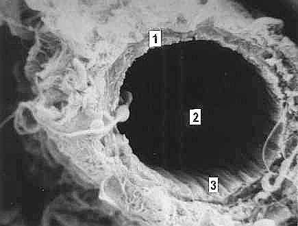 |
| Figure 1. Blood capillary in the normal testis of the control rat. (1) capillary wall, (2) lumen, (3) endothelial layer. |
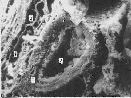 |
| Figure 2. Blood capillary after cadmium administration. The capillary wall (1) is damaged. Near the capillary the vacuoles (3) are visible. Lumen of the blood capillary (2). |
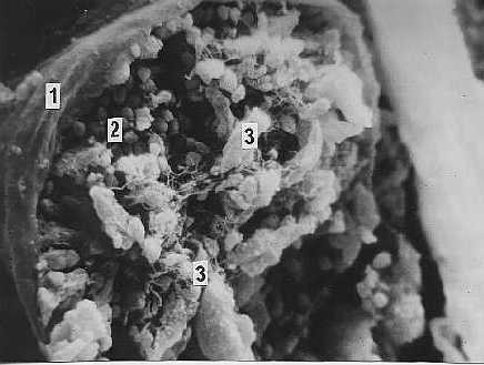 |
| Figure 3. Seminiferous tubule in the testis of the rat after cadmium administration contains necrotizing germinal cells (2), which evacuate into the lumen of the tubule (3). Lamina propria (1). |
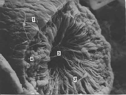 |
| Figure 4. Seminiferous tubule of the control rat. View of the lamina propria from the surface area (1). Spermatozoa (2) release into the lumen (3) during proceeding spermatogenesis. The youngest developmental stages of the germinal cells, spermatogonia and primary spermatocytes (4) located on the periphery of the tubule. |
|
|
|
|
|
|
|
[Toxicology] |
[Pathology] |
[Reproduction Sciences] |