Poster
# 25
![]()
Poster |
|
6th Internet World Congress for Biomedical Sciences |
María Jesús Ramírez-Expósito(1), José Manuel Martínez-Martos(2)
(1)Unit of Physiology. University of Jaen - Jaén. Spain
(2)Unit of Physiology. University of Jaén - Jaén. Spain
|
|
|
|
|
|
|
[Cell Biology & Cytology] |
[Neuroscience] |
[Physiology] |
 |
| Figure 1. Semithin sections showing the total thickness of frontal cortex. |
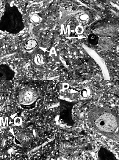 |
| Figura 2. Light microscopy image of toluidine blue stained semithin section of the frontal cortex of rats showing astrocytes (A) and microglia-oligodendrocytes (M-O). |
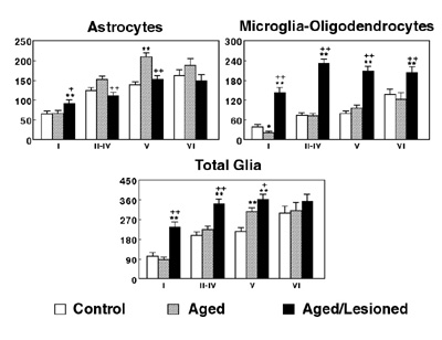 |
| Figure 3. Glial density (Mean±SEN) expressed as the number of astrocytes (A), microglia-oligodendrocyte (MO) and total glia (TG) per 106 µm3 of tissue in the diferent cortical layeror control, old and old and lesioned groups (** denotes statistical significance, p<0.01, between young and the other groups. + and ++ denote statistical significance, p<0.05 and p<0.01 respectively, between old and old and lesioned groups). |
|
|
|
|
|
|
|
[Cell Biology & Cytology] |
[Neuroscience] |
[Physiology] |