Poster
# 15
![]()
Poster |
|
6th Internet World Congress for Biomedical Sciences |
Jesus Gonzalez Garcia(1), Marcial García Rojo(2), Francisco Martín Dávila(3), Rafael López Pérez(4), Margarita Delgado Portela(5), Manuel Carbajo Vicente(6)
(1)(2)(3)(4)(5)(6)Servicio de Anatomía Patológica. Complejo Hospitalario de Ciudad Real - Ciudad Real. Spain
|
|
|
| |
|
[Orthopedics & Traumatology] |
[Pathology] |
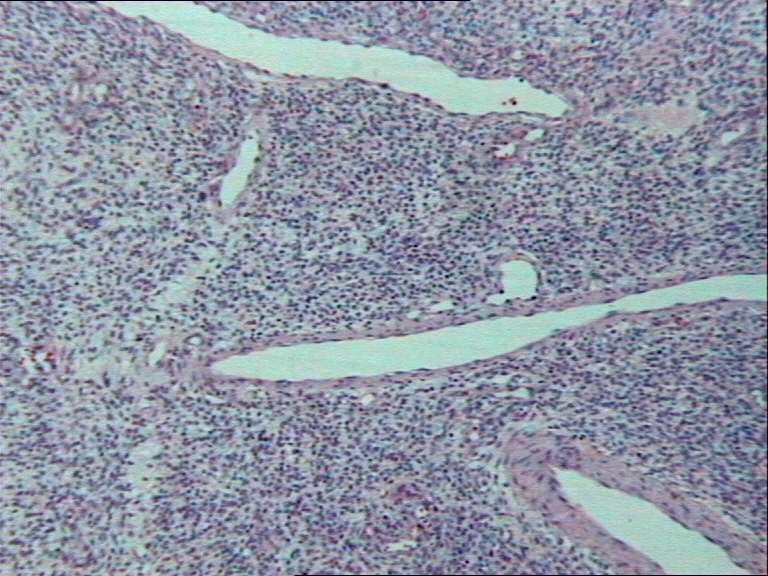 |
| Fig. 6: Higher vascularized areas. |
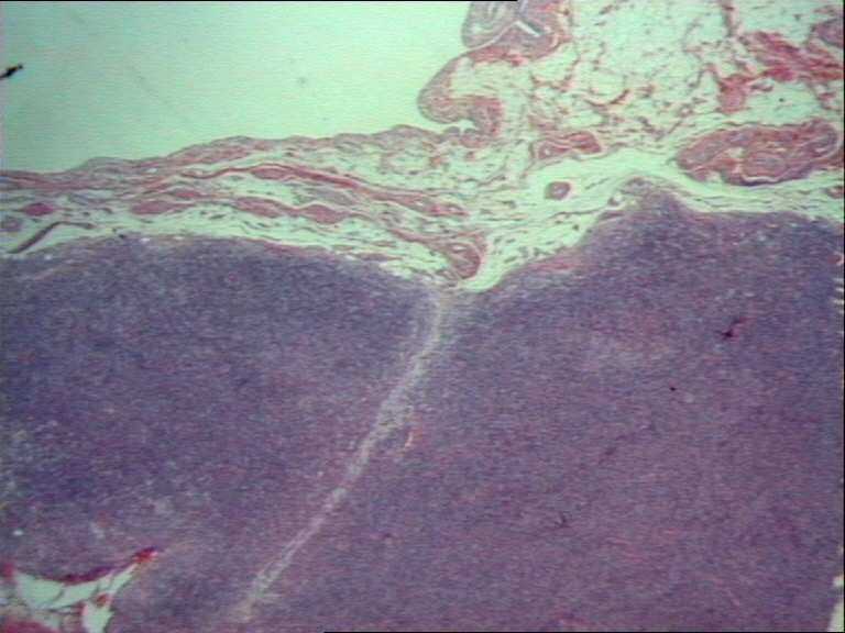 |
| Fig. 7-A: Well delimited boundaries under synovyum. |
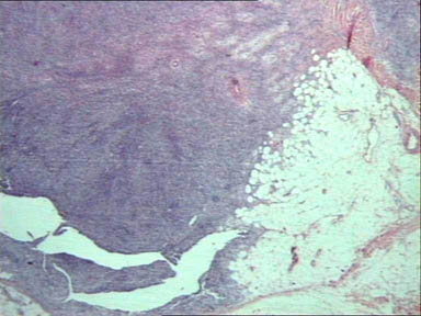 |
| Fig. 7-B: More blurred and irregular boundaries between neoplasm and adjacent fatty tissue. |
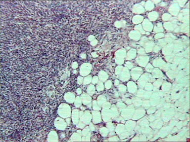 |
| Fig. 7-C: More blurred and irregular boundaries between neoplasm and adjacent fatty tissue. |
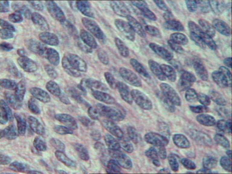 |
| Fig. 8-A: High power view of monomorphic fusiform cells, with bad delimited citoplasmic limits, slight nuclear atypia and inconspicuous nucleolus. |
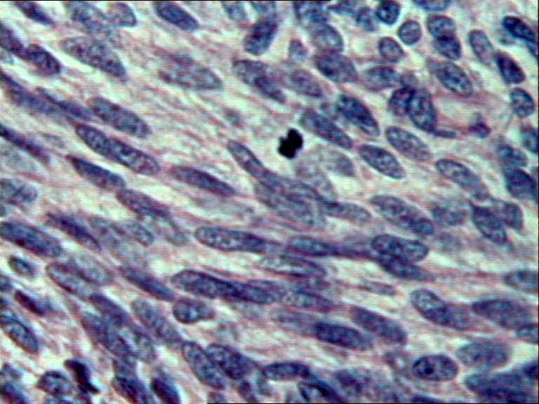 |
| Fig. 8-B: Mitotic figures. |
|
|
|
| |
|
[Orthopedics & Traumatology] |
[Pathology] |