Poster
# 8
![]()
Poster |
|
6th Internet World Congress for Biomedical Sciences |
Francisco Martín Dávila(1), Marcial García Rojo(2), Jesus Gonzalez Garcia(3), Margarita Delgado Portela(4), Rafael López Pérez(5), Manuel Carbajo Vicente(6)
(1)(2)(3)(4)(5)(6)Servicio de Anatomía Patológica. Complejo Hospitalario de Ciudad Real - Ciudad Real. Spain
|
|
| |||
|
[Obstetrics & Gynecology] |
[Oncology] |
[Pathology] |
 |
|
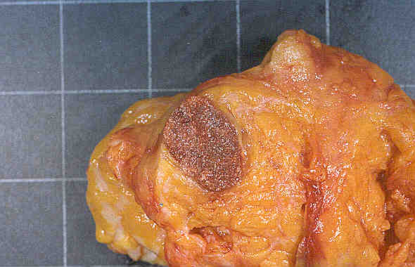
Figure 1. Macroscopically, two mammary fragments each of them with a nodular tumor of 2,8x2x1,7 cm. and 1 cm. They were very well demarcated from adjacent fatty tissue with a shinny brown cut surface. |
 |
|
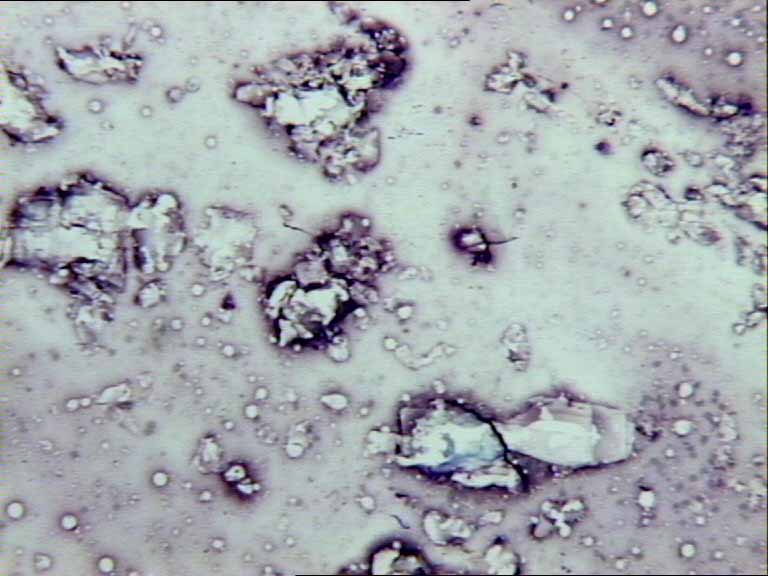
Figure 2. Imprints show crystals with irregular and many times trapezoidal contours negative under polarized light and scattered macrophages. |
 |
|
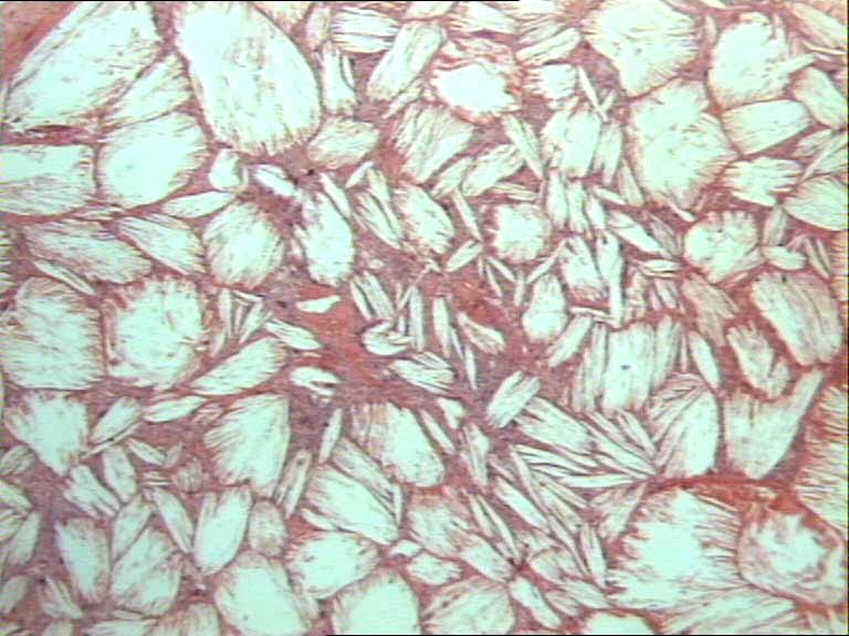
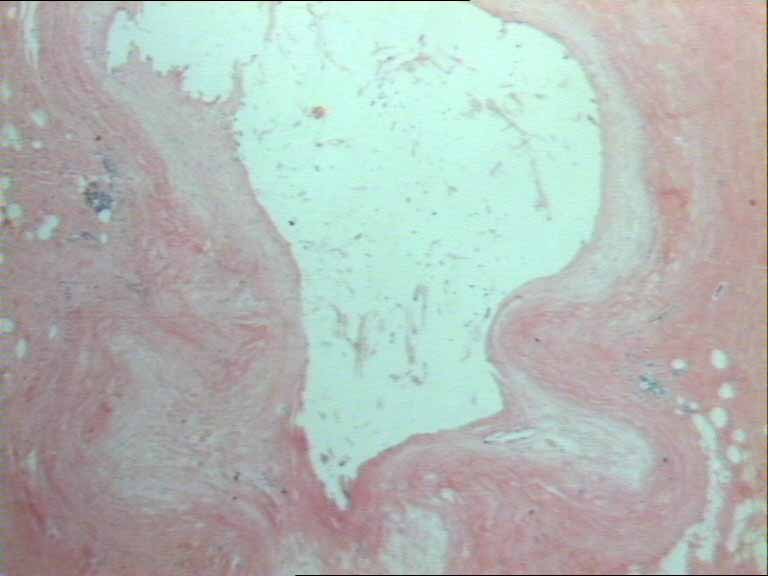
Figure 3. On histologic sections, many large and irregular aggregates of tightly packed cholesterol needle-like crystals in parallel or radial arrays were observed in both tumors. |
 |
|
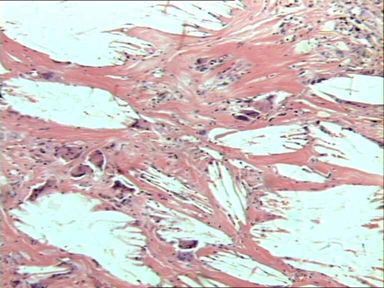
Figure 4. Collagen bands containing macrophages and foreign-body giant cells were noted between crystal aggregates. |
 |
|
Figure 5. In adjacent mammary tissue there were distended ectatic ducts containing foamy histocytes, calcium deposits and immersed in a fibrous stroma. |
|
|
| |||
|
[Obstetrics & Gynecology] |
[Oncology] |
[Pathology] |