Comunicación
      Nº 071

IICVHAPp.gif (9537 bytes)

IndiceIndice

TUMOR NEUROEPITELIAL DISEMBRIOPLÁSICO.
ESTUDIO CLÍNICO, HISTOLÓGICO E INMUNOHISTOQUÍMICO CUATRO NUEVOS CASOS

Teresa Ribas Ariño, Elena García Lagarto, Francisco Miguel Izquierdo García, Santos Salas Valién, Ángela Miguelez Simón.

Comentarios

[TITULO] [INTRODUCCIÓN] [MATERIAL Y MÉTODOS] [RESULTADOS] [ICONOGRAFÍA] [ICONOGRAFÍA-2] [DISCUSIÓN] [BIBLIOGRAFÍA]

RESULTADOS

ICONOGRAFÍA-2


ICONOGRAFÍA

fig. 1
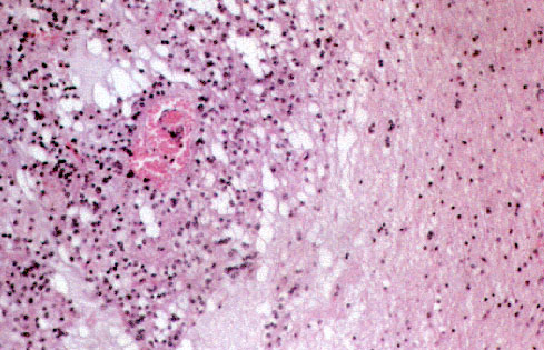
Fig.1: Zona de transición con el cortex circundante, con buena demarcación entre el mismo y el tumor. H&E x20.


fig. 2
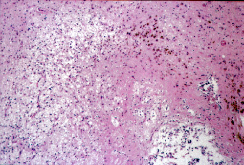
Fig.2: Celularidad monomorfa y hábito multinodular. H&Ex10.


fig. 3
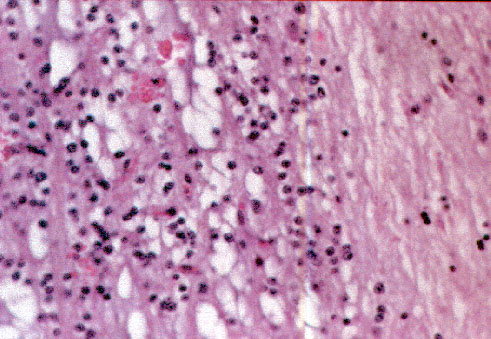
Fig.3: Células similares a oligodendrocitos, monomorfas en patrón difuso y laxo. H&Ex40


fig. 4
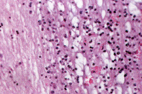
Fig.4: Disposición de las células en hileras paralelas a l cortex. H&Ex40.


[TITULO] [INTRODUCCIÓN] [MATERIAL Y MÉTODOS] [RESULTADOS] [ICONOGRAFÍA] [ICONOGRAFÍA-2] [DISCUSIÓN] [BIBLIOGRAFÍA]

RESULTADOS

ICONOGRAFÍA-2


Teresa Ribas Ariño, Elena García Lagarto, Francisco Miguel Izquierdo García, Santos Salas Valién, Ángela Miguelez Simón.
Copyright © 1998. Reservados todos los derechos.