Presentation
# 3
![]()
Presentation |
|
6th Internet World Congress for Biomedical Sciences |
Albert Sun(1), Bozena Draczynska-Lusiak(2), Grace Sun(3)
(1)(2)(3)Department of Pharmacology. University of Missouri - Columbia. United States
|
|
|
|
 |
|
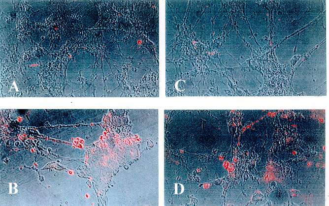
Fig 1. Uptake of brain lipoprotein (BLP) labeled with fluorescent Di-I by primary cortical neurons in culture. Brain LPs were prepared using lipids extracted from rat brain cerebral cortex and subjected to oxidation as described in text. Native and oxidized BLP enriched with apoE3 or apoE4 were incubated with fluorescent Di-I. The Di-I labeled BLPs were added to primary cortical neurons and incubated for 4 hrs. The cells were washed and fixed with 3% formaline in phosphate buffer, pH 7.4. Confocal and fluorescence microscopy was performed with epifluorescent illumination using the rhodamine filter package. The four panels represent each of the following treatments: Panel A: Cells treated with Di-I labeled native BLP enriched with apoE3; Panel B: Cells treated with Di-I labeled oxidized BLP enriched with apoE3; Panel C: Cells treated with Di-I labeled native BLP enriched with apoE4; and Panel D: cells treated with Di-I labeled oxidized BLP enriched with apoE4.
|
 |
| Fig 2. Time course of uptake of BLPs labeled with |
 |
|
Fig. 3. Effect of native or oxidized BLPs on cell viability as assessed by MTT reduction assay. Cells were treated with native (naBLP) or oxidized BLP (oxBLP) enriched with apoE3 for 48 hrs and were subsequently harvested for MTT assay as described in Methods. In addition, cells were treated with 0.5 mM of resveratrol (R) in order to examine whether oxidative stress is involved in cell death caused by oxBLPs. Data are mean +/- SD from 4 dishes.
|
 |
|
Fig. 4. Oxidized BLPs induce cell death as assessed by lactate dehydrogenase (LDH) release assay.
The treatment conditions were the same as described in Fig 3 except that the incubation media were recovered at the end of incubation time and the clear centrifuged media were used for LDH assay as described in the Methods. Data are mean +/- SD from 4 dishes.
|
 |
|
Fig. 5. Effects of Aß and BLP in survivability of primary cortical neurons. Cells were treated with native or oxidized BLP enriched with apoE4 as described in Fig 3 except that incubation time was 24 hrs and cells were harvested for MTT assay. Aß (2µM) and FeCl2+/DTP (50 µM ) were added to the medium at the beginning of incubation. Data represent mean +/- SD from 4 dishes.
|
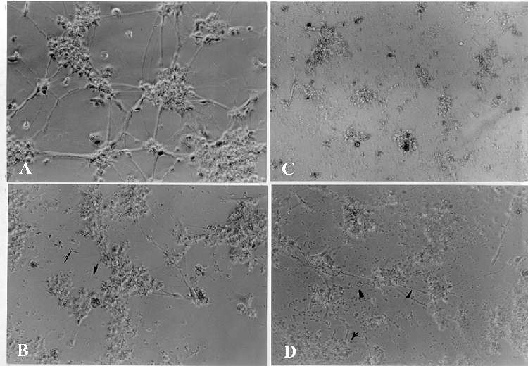 |
| Fig. 6. Morphological changes of the cells after Aß and/or oxidized BLP treatment. Primary neurons in culture were exposed to Aß (2µM) and/or oxidized BLP enriched with apoE4 for 24 hr and examined under phase contrast microscope (Nikon Diaphot 300, Nikon Corp. Melville, NY). A: Control cells without treatment; B: Cells treated with Aß alone; C: Cells treated with Aß together with oxidized BLP; D: Cells treated with oxidized BLP alone. |
|
|
|
|