Poster
# 108
![]()
Poster |
|
6th Internet World Congress for Biomedical Sciences |
Johan Van Doornik(1), Dimitris Patikas(2), Daniel Olivier(3), Gabriella Cerri(4), Michel Ladouceur(5), Jens Bo Nielsen(6), Thomas Sinkjaer(7)
(1)Centre for Sensory-Motor Interaction - Aalborg. Denmark
(2)Aristotle University of Thessaloniki - Thessaloniki. Greece
(5)Dept. of Medical Informatics and Image Analysis. Center for Sensory-Motor Interaction (SMI). Institute of Electronic Systems, Aalborg University - Aalborg O. Denmark
(7)Center for Sensory Motor Interaction. Aalborg University - Aalborg. Denmark
|
|
[Neuroscience] |
[Physiology] |
[Neurology] |
[Physical Therapeutics & Rehabilitation] |
|
Preliminary stretch reflex experiments
Figure 1 shows the TA EMG, SOL EMG, ankle torque, and ankle angle at three different stretch velocities from participant 1. The TA EMG shows the M1, M2 and M3 peak. The SOL EMG remains very small during the stretch. The peak values M1, M2 and M3 from this and three other participants are measured at six different stretch velocities and are shown in figure 2. These results indicate that the M3 component is modulated by the stretch velocity and appears to be maximal for 50 deg/s. This stretch velocity will be used for the following experiments.
Balance training experiments
In figure 3 the TA EMG, SOL EMG, Ankle torque, and ankle angle during the TA stretch reflex are shown from before and after the balance training of participant 5. Surprisingly, the M3 component shows a decrease, whereas the M1 component remains the same. Figure 4 shows the MEP in the SOL and TA at 35%, 55%, 75% and 95% stimulus intensity before and after the balance training in the same participant. Figure 5 shows the peak-to-peak value of the MEP as a function of the stimulus intensity (the input-output curve). The reduction of the plateau phase of the fitted Boltzmann sigmoidal curve is significant within a 95% confidence interval.
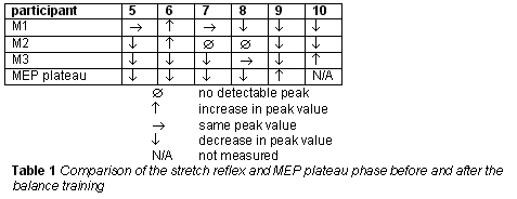
Table 1 shows the results for 6 participants. Indicated is whether the stretch reflex components increased, decreased or remained the same, and whether the plateau phase of the MEP increased or decreased within a 95% confidence interval
|
|
[Neuroscience] |
[Physiology] |
[Neurology] |
[Physical Therapeutics & Rehabilitation] |
|