Poster
# 80
![]()
Poster |
|
6th Internet World Congress for Biomedical Sciences |
Alberto Pizarro Gallardo(1)
(1)Facultad de Medicina U.A.N. - Tepic. Mexico
|
|
|
|
|
|
[Neurology] |
[Pathology] |
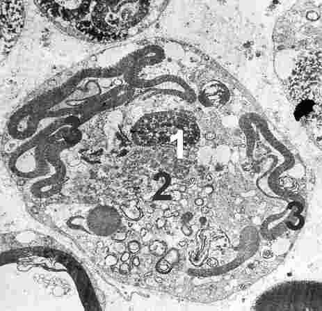 |
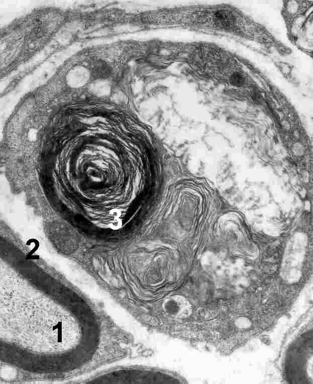
| Fig. 1. Altered Schwann cell . There is necrosis of the cell of Schwann. 1:nucleus, 2:mitochondria, 3 : degenerative myelin . 15,000 X |
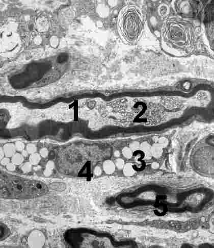 |
| Fig. 2. Schwann cell. 1: normal axon 2:small mitochondria, 3: inclusion,4: nucleus, 5: normal myelin, 5: demylination. 12,000 X |
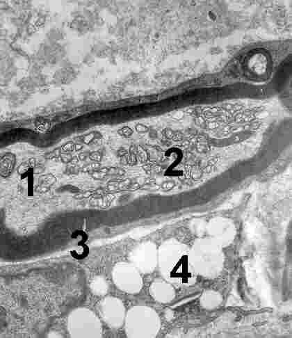 |
| Fig. 3. Schwann cell with fat inclussions. 1: axon 2: small mitochondria, 3: normal myelin, 4:inclusion, 20,000 X |
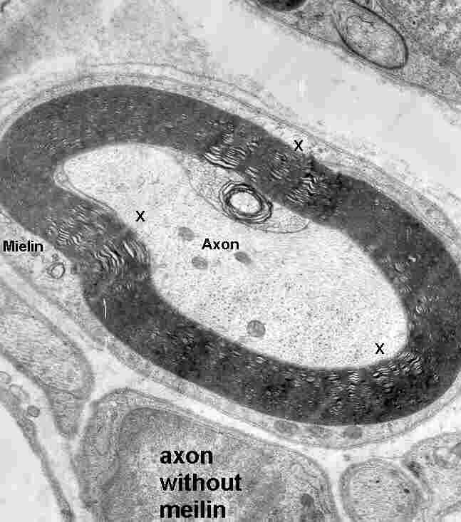 |
| Fig. 4. Schwann cell. Axon myelin, x: demylination areas 25,000 X |
 |
| Fig. 5. normal axon, 2: normal myelin, 3: demylination 4: inclusion. 25,000x |
|
|
|
|
|
|
[Neurology] |
[Pathology] |