Poster
# 69
![]()
Poster |
|
6th Internet World Congress for Biomedical Sciences |
Stephan Lolov(1), Dinka Petrova(2), Stanimir Kyurkchiev(3), George Edrev(4)
(1)Institute of Biology and Immunology of Reproduction. Bulgarian Academy of Sciences - Sofia. Bulgaria
(2)(3)Institute of Biology and Immunology of Reproduction; Bulg. Acad. Sci. - Sofia. Bulgaria
(4)Dept. Otorhinolaryngology. Transport Medical Institute - Sofia. Bulgaria
|
|
|
|
|
|
[Immunology] |
[Otorhinolaryngology] |
Abstract
Cholesteatoma is a destructive lesion of the human middle ear with unknown etiopathogenesis. Immunohistochemical assays with specific monoclonal antibodies have been used to register expression and distribution of several well-known antigens. In this study, the authors have looked for abnormal or unknown cholesteatomas constituents generating monoclonal antibodies.
BALB/c mice were immunized with subcutaneous implantation of the cholesteatomas sac and matrix. Hybridomas have been generated following procedures known in the art. The screening for clones with specific antibodies production was done using enzyme linked immunosorbend assay (ELISA) against saline tissue extract and immunofluorescence (IF) with paraffin embedded tissue sections.
The monoclonal antibody designated 1F8 showed positive reaction in ELISA and in IF when tested against both cholesteatoma and ear canal skin. At the same time when supernatants from different cell and tissue cultures have been used as an antigen in ELISA, 1F8 reacted only with cholesteatomas supernatant. In Western blot it recognizes three small bands and a prominent one, with MW of approximately 70 kD. Using immunohistochemistry the antigen was localized in the subepithelial connective tissue fibers of the skin and the cholesteatoma. The antigen of IF8 was detected in all tested cholesteatomas. Studies are now in progress to characterize the antigen in more details.
Key words:
cholesteatoma; monoclonal antibodies; immunohistochemistry
Introduction
Cholesteatoma is a destructive lesion of the human middle ear with unknown etiopathogenesis. The process of its formation is an object of intensive research and several controversial theories have been formulated:
Immunohistochemical assays with specific monoclonal antibodies have been used to register expression and distribution of several well-known antigens (fibronectin 4, 20, 24, interleukin (IL)-1 alpha and IL-2 1, 8, 10, 21, 22, epidermal growth factor, transforming growth factor-beta 1 and 2 23, tumor necrosis factor-alpha 15, 25, keratinocyte growth factor 7, 11, c-myc protein 6, different cell surface peptidases, parathyroid hormone-related protein 5 and many others), which may modulate cell growth and differentiation, as well as invasion of healthy tissues and bone resorption. The existence of a defect in the normal biology and/or biochemistry of cholesteatomas constituents have been suggested, but nothing specific for this entity have been reported.
In this study, we have looked for abnormal or unknown cholesteatomas constituents generating monoclonal antibodies.
Materials and methods
Some of the methods used were based on the classical protocols with just minor modifications:
|
SDS Gel Electrophoresis |
|
|
Protein Concentration Determination |
|
|
Silver Staining of Gels |
|
|
Electroblotting of SDS-PAA gels |
|
|
Thawing /Freezing Hybridomas and Myelomas |
|
|
Cloning by Limiting Dilution |
|
|
Ascites Production |
Immunization
The mice were given an anaesthetic dose of 0.01 % Halothanum thymolo (Spofa) and immobilized on surgical board. After removal of the fur by shaving and dampening with ethanol, a V-shaped cutaneous incision was made in the dorsal region. About 1 mm3 piece of xenogenous tissue, handled with forceps was inserted through a small slit in the animals subcutaneous tissue until becomes completely embedded. After that, the skin was carefully sutured.
Two and four weeks later, the procedure of tissue implantation was repeated, each time with a histologicaly proved cholesteatoma, from a different patient. Three days before the fusion, a final boost was done intraperitonealy with saline extract of cholesteatoma (10 m g total protein).
Saline tissue extract
As soon as possible after excision, cholesteatomas tissue and skin from external meatus of seven patients were minced and cells were disrupted with ten cycles of freezing to -20oC and thawing to room temperature in the presence of PBS with PMSF, aprotinin and trypsin inhibitor. After brief spin at 1850 G the supernatants were collected and stored at -20oC until used.
Fusion
P3X63-Ag8.653 cells (non secreting, 8-azaguanine resistant, HPRT myeloma; 2.5 x 10⁷ cells) were added to spleen cells (9.2 x10⁷) from the immunized BALB/c mice. PEG/DMSO solution was added under agitation folowing well established protocol (Peter MacCallum). The cell pellet was resuspended in RPMI 1640 medium supplemented with 10% fetal calf serum and plated into five tissue culture plates and incubated in the presence of hypoxanthine, aminopterin, and thymidine medium in a humidified 37oC, 5% CO2 incubator.
Screening
First clones were seen after 7-10 days. The first screening procedure was started after 2 weeks, with a second and third, a few days later. Only wells with the confirmed presence of one or two big clones, were tested for the secretion of desired antibody. Both enzyme linked immunosorbend assay (ELISA) and indirect immunofluorescence (IF) were used as screening procedures.
Hybridomas secreting specific antibodies were passed through limiting dilution and retested until all wells were considered positive.
ELISA
Immunohistochemistry
Fluorescence visualization:
Peroxidase visualization (Avidin-biotin affinity method, ABA):
Western blot
All steps were done with constant shaking.
Photomicrography
Kodak Technical Pan was the film of choice for Western blot documentation. It was processed to give high contrast. Thus the small differences were accentuated.
Color negative film (AGFA HDC 100) was developed and printed commercially. To avoid the loss of control over the printing process and to receive comparable results:
Results and discussion
It should be kept in mind that some routine immunization techniques involve the risk of causing conformational changes to the antigen during its purification, cross-linking with carrier or when emulsified in an adjuvant. This can result in the production of antibodies against inactive, denatured antigens. In the present study a new immunization approach was used - the whole cholesteomas tissue (sac and matrix) was deposited several times into the subcutaneous tissue of experimental animal. This technique provides an easy way to immunize animals with an uncharacterized and unlocalized antigen in a native conformation. At the same time, xenogenous tissue implantation would result in a prolonged presence of antigen, which would lead to an augmentation of the immune response. So, the use of adjuvant with all its side effects like sterile abscesses, granuloma formation and unwanted antibodies generation is avoided by the present approach.
|
A |
B |
|
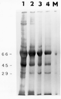
Silver-stained gel of a 10% SDS-PAGE of saline tissue extract from cholesteatoma (A) and ear canal skin (B). M-lanes contained protein standards with their sizes listed in kilodaltons. |
There were no significant differences between cholesteatoma and skin, when several concentrations were compared under reducing conditions (lanes 1 - 1 µg total protein; lanes 2 - 0.5 µg protein; lanes 3 - 0.25 µg protein; lanes 4 - 0.125 µg protein). |
|
|
|
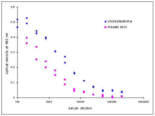
Just before the booster immunization a test bleeding was performed and antibody titers were assayed by ELISA. The sigmoid curves obtained by testing serial dilutions of mouse serum demonstrated stronger polyclonal response against cholesteatomas extract. |
|
A |
B |
|
C |
D |
|
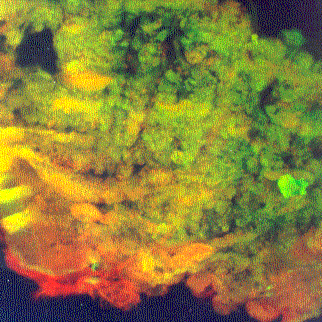
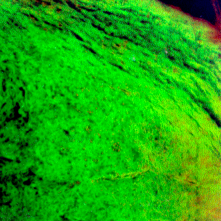
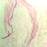
Indirect immunofluorescence of ear canal skin (A) and cholesteatoma (B) after incubation with cell culturing supernatant from the hybridoma 1F8. |
The negative controls of skin (C) and cholesteatoma (D) were treated with supernatant from myeloma cell line. |
|
Hematoxilin-eosin staining of cholesteatoma with cholesterol clefts. |
|
|
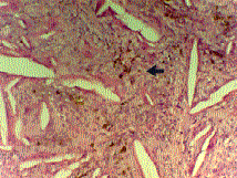
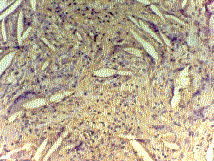
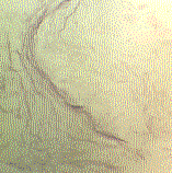
There were no specific reaction when serial sections were incubated with supernatant from myeloma cell line (negative control). Brown nodules (arrow) represented not color reaction product, but haemosiderin. |
|
|
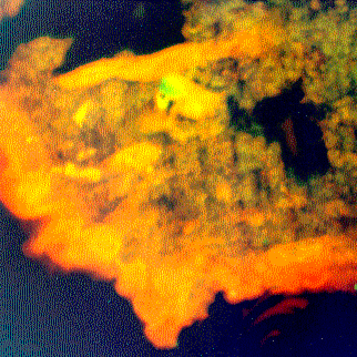
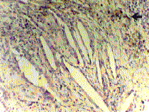
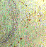
The serial sections, after incubation with culture supernatant from hybridoma 1F8, demonstrated specific brown staining of the connective tissue. |
|
|
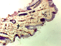
When the skin from external meatus was used as an antigen in immunohistochemistry, there was uniform specific staining in the subepithelial space (thin arrow) and more fibrous in deeper layers (thick arrow). |
|
|
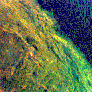
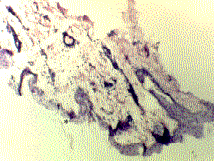
The serial section was incubated with supernatant from myeloma cell line and used as negative control. Some darkly stained areas of epithelium (asterisk), observed also in previous picture, represented staining artifacts. |
|
|
Hematoxilin-eosin stained squames of dead keratin in an acquired cholesteatoma. |
|
|
The serial paraffin section, incubated with antibody 1F8, followed by visualization with ABA method. Note the brown stained granular structures (specific reaction) and blue dyed squames (hematoxilin counterstaining). |
|
|
The negative control was incubated with supernatant from myeloma cell line and subjected to ABA procedure. |
|
|
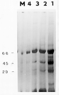
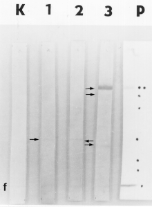
Evaluation of the specificity of monoclonal antibody 1F8 (lane 3) and some others (BX - lane 1 and BY - lane 2) by Western blot analyses using 12% PAA gel and cholesteatomas saline extract (100 µg /lane) under denaturing conditions. The lane K was incubated with supernatant from myeloma cell line and used as negative control. To verify transfer efficiency, Ponceau S staining was performed (lane P). The dye front (f) was also marked. 1F8 recognizes three small bands and a prominent one, with MW of approximately 70 kD. |
|
|
|
|
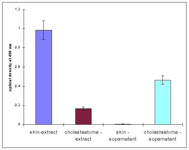
When supernatants from different cell cultures (U937, Jurkat, P3U1, HUVEC and SK MEL1 - data not shown) and tissue cultures have been used as an antigen in ELISA, 1F8 reacted only with cholesteatomas supernatant. At the same time when saline tissue extracts were absorbed on the PVC plate, the reaction against skin was stronger. |
We succeeded to detect the antigen of 1F8 in all cholesteatomas cases (seven) studied.
Conclusions
The data of the present work suggest that generation of monoclonal antibodies using tissue implantation as an immunization procedure is a promising approach to obtain an immunological tool for unknown antigen isolation and characterization. Unfortunately, we failed to detect cholesteatomas specific antigen. Other immunization and fusion experiments are now in progress.
The monoclonal antibody designated 1F8 showed positive reaction in ELISA and in IF when tested against both cholesteatoma and ear canal skin. It recognizes several bands in Western blot after SDS-PAGE. The antigen of 1F8 was detected in all tested cholesteatomas. It is expressed only in skin and cholesteatoma (data not shown) and possesses some interesting characteristics.
References
|
|
|
|
|
|
[Immunology] |
[Otorhinolaryngology] |