Poster
# 15
![]()
Poster |
|
6th Internet World Congress for Biomedical Sciences |
Jesus Gonzalez Garcia(1), Marcial García Rojo(2), Francisco Martín Dávila(3), Rafael López Pérez(4), Margarita Delgado Portela(5), Manuel Carbajo Vicente(6)
(1)(2)(3)(4)(5)(6)Servicio de Anatomía Patológica. Complejo Hospitalario de Ciudad Real - Ciudad Real. Spain
|
|
|
| |
|
[Orthopedics & Traumatology] |
[Pathology] |
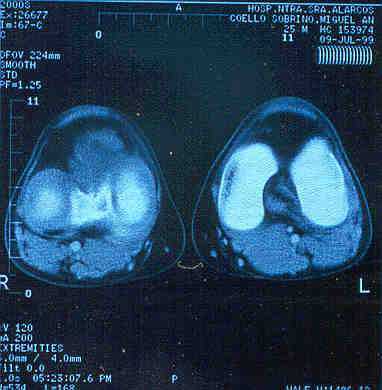 |
| Fig. 1: TAC image: Mass in anterior compartment of the right knee, in close relation with intercondyleal space and inner meniscus. |
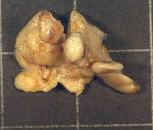 |
| Fig. 2: Macroscopic view of the tumor with a polilobulated and something villous surface. |
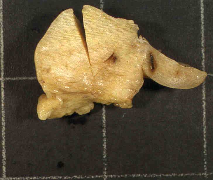 |
| Fig. 3: Homogeneous and well-delimited neoplasm on cut surface. |
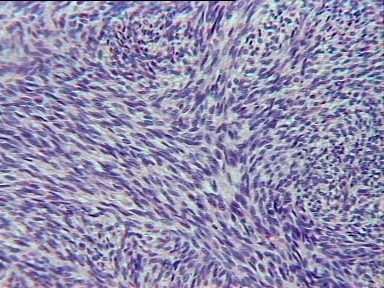 |
| Fig. 4-A: High density cellular areas with a lightly fasciculated appearance. |
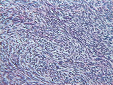 |
| Fig. 4-B: High density cellular areas with a lightly fasciculated appearance. |
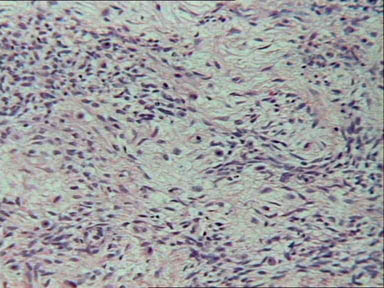 |
| Fig. 5-A: Loose cellular and myxoid areas with low and high power view. |
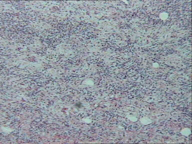 |
| Fig. 5-B: Loose cellular and myxoid areas with low and high power view. |
|
|
|
| |
|
[Orthopedics & Traumatology] |
[Pathology] |