Comunicaciµn
N¤ 041

Comunicaciµn |
|
Giorgio Gherardi, M.D., Cristina Marveggio, B.D., Stefania Rossi, M.D.
![]()
|
|
 |
|
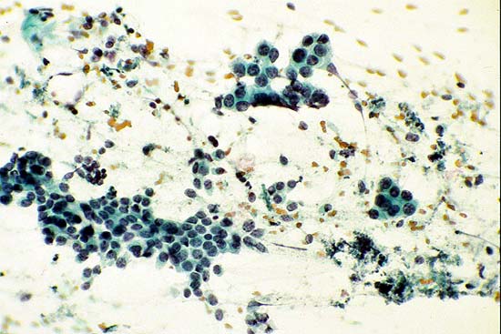
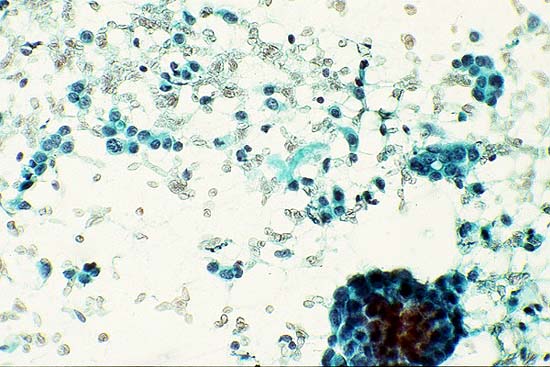
Figure 1A: Case 12. Palpable florid hyperplasia. The aspirate cytologic score was 3+3+3+2+2+3=16 (Papanicolaou stain, x500). DNA-image analysis of the aspirate demonstrated a SPD histogram. |
 |
|
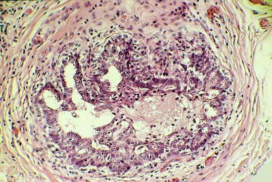
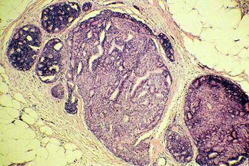
Figure 1B: Case 12. Palpable florid hyperplasia. Histologic view of ductal hyperplasia without atypia (Hematoxylin-eosin, x250). |
 |
|
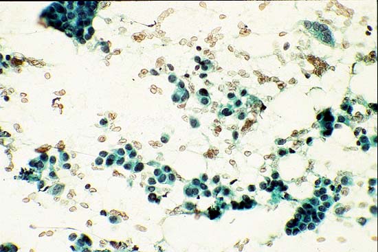
Figure 2A: Case 3. Palpable 1.8-cm superficial nodule. The aspirate cytologic score was 3+2+3+2+2+3=15. (Papanicolaou stain, x400). DNA-image analysis of the aspirate demonstrated a SPD histogram. |
 |
|
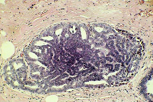
Figure 2B: Case 3. Palpable 1.8-cm superficial nodule. Histologic view of ductal hyperplasia with atypia (hematoxylin-eosin, x180). |
 |
|
Figure 3A: Case 18. Palpable 1.2-cm superficial lesion. The aspirate cytologic score was 3+2+3+3+2+3=16. (Papanicolaou stain, x600). DNA-image analysis demonstrated a aneuploid histogram with DI = 1.42. |
 |
|
Figure 3B: Case 18. Palpable 1.2-cm superficial lesion. Histologic section shows a ductal carcinoma in situ with cribriform and papillary patterns (hematoxylin-eosin x80). |
|
|