
 Virtual slide # 2. Slow growing scalp mass in 18 year-old male
|
|
Mirta Garcia Jardón*, Lech Banach**, Andrew Stepien**, Irena Targonska**, Charl Hobson*** |
||
|
18 year-old male presented with one year history of a slow growing mass on the scalp. CT-scan showed a soft tissue tumour infiltrating skull penetrating intracranially (figure 1). The growth was located in the centre of the parietal region. Excision of the mass, including bone particles was done. The specimen was sent to the Department of Pathology for histological diagnosis.
Two surgical specimens were received at the Department. The smallest was made up of small soft tissue fragments admixed with bone particles; and the tumour itself (no picture of the smallest shown). The macroscopic appearance of the tumoral mass is seen in figures 2 and 3.
Se muestran los resultados de biopsia en un paciente con una masa tumoral de partes blandas del cuero cabelludo, de crecimiento lento, de alrededor de un año de duración, que fué resecada en un varón de 18 años. El tumor era clínicamente blando, no doloroso, fijado al cráneo. La TAC de cráneo mostró infiltración del hueso, con penetración en la cavidad craneana. Se realizó biopsia excisional y se envió para estudio a nuestro departamento. Este trabajo presenta los hallazgos clínico-morfológicos (macro y microscópicos) de la biopsia y se realizan algunas consideraciones acerca de su posible histogénesis.
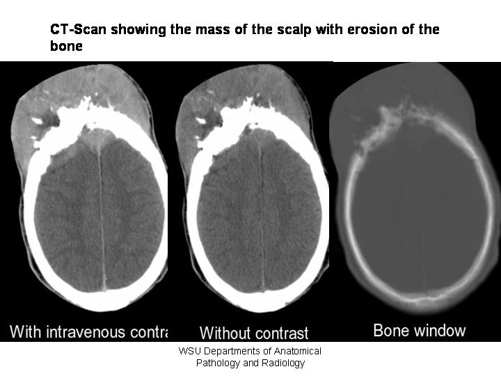 fiogf49gjkf0dFigure 1: CT-Scan of the soft tissue mass showing the erosion of the bone. The mass measured 99x47mm with bone destruction. Right side view shows intracranial extention of 64x17x50mm. Brain tissue and ventricular system are preserved. No signs of compression noted.">
Figure 1: CT-Scan of the soft tissue mass showing the erosion of the bone. The mass measured 99x47mm with bone destruction. Right side view shows intracranial extention of 64x17x50mm. Brain tissue and ventricular system are preserved. No signs of compression noted. fiogf49gjkf0dFigure 1: CT-Scan of the soft tissue mass showing the erosion of the bone. The mass measured 99x47mm with bone destruction. Right side view shows intracranial extention of 64x17x50mm. Brain tissue and ventricular system are preserved. No signs of compression noted.">
Figure 1: CT-Scan of the soft tissue mass showing the erosion of the bone. The mass measured 99x47mm with bone destruction. Right side view shows intracranial extention of 64x17x50mm. Brain tissue and ventricular system are preserved. No signs of compression noted.
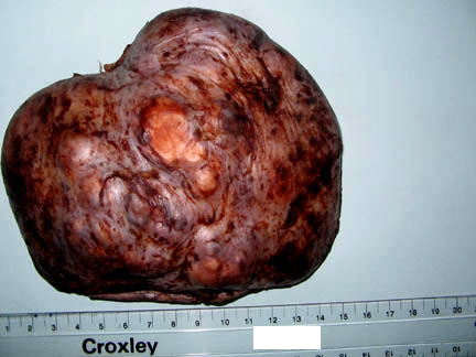 fiogf49gjkf0dFigure 2: Macroscopic appearance of the resected mass. Irregular, polylobulated appearance of the tumor, covered by skin is seen.">
Figure 2: Macroscopic appearance of the resected mass. Irregular, polylobulated appearance of the tumor, covered by skin is seen. fiogf49gjkf0dFigure 2: Macroscopic appearance of the resected mass. Irregular, polylobulated appearance of the tumor, covered by skin is seen.">
Figure 2: Macroscopic appearance of the resected mass. Irregular, polylobulated appearance of the tumor, covered by skin is seen.
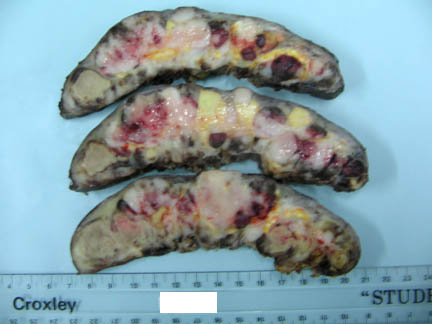 fiogf49gjkf0dFigure 3: Sectional surface of the tumour. Multinodular appearance of the tumour, with some haemorrhagic nodules, whitish, yellowish or sand-like due to necrosis.">
Figure 3: Sectional surface of the tumour. Multinodular appearance of the tumour, with some haemorrhagic nodules, whitish, yellowish or sand-like due to necrosis. fiogf49gjkf0dFigure 3: Sectional surface of the tumour. Multinodular appearance of the tumour, with some haemorrhagic nodules, whitish, yellowish or sand-like due to necrosis.">
Figure 3: Sectional surface of the tumour. Multinodular appearance of the tumour, with some haemorrhagic nodules, whitish, yellowish or sand-like due to necrosis.
|
||
|
|
Web mantenido y actualizado por el Servicio de informática uclm. Modificado: 16/06/2015 17:26:32