

|
|
A Forteza*, Alexandra Gené*, R Ramos*, F Terrasa*, M Company*, Ramón Canet* |
||
|
A female fetus, stillborn at 27 weeks gestation, who presented a generalized subcutaneous edema without other malformations.
MACROSCOPIC FINDINGS
In the macroscopic exam we found in the thoracic cavity a pleural effusion and a big growing expansive mediastinal neoplasm that measured 8 cm and weighted 56 g. The cut surface showed fleshy appearance and some cystic areas. The lungs were small, weighted 10 g but had a correct lobulation.
|
||
|
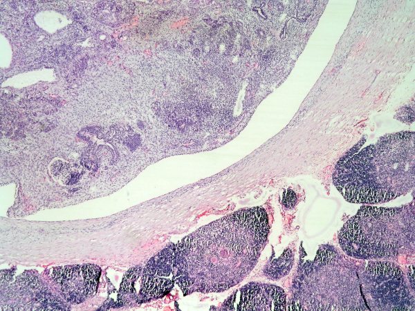
Preparación amablemente digitalizada por los responsables de WebMicroscope (Finlandia) H&E Lung | |||
|
|
||
|
ANTERIOR MEDIASTINAL IMMATURE TERATOMA
|
||
|
MICROSCOPIC FINDINGS The mediastinal neoplasm was composed of immature and mature tissues representing each of the three layers derived from the embryonic disk. We found dermal structures and neural, gastrointestinal, bronchial, muscular and fatty tissues with some cystic lesions. We observed some immature neuroectodermic areas with neuroepithelial cells forming tubules and rosettes. Surrounding the neoplasm atrophic thymic rests could be found. DIAGNOSIS: 1. HYDROPS FETALIS. 2. ANTERIOR MEDIASTINAL IMMATURE TERATOMA 3. BILATERAL PULMONARY HYPOPLASIA CAUSED BY INTRATHORACIC COMPRESSION
.jpg) fiogf49gjkf0d"> fiogf49gjkf0d">
 fiogf49gjkf0d"> fiogf49gjkf0d">
|
||
|
1 - Froberg MK, Brown RE, Maylock J, Poling E. In utero development of a mediastinal teratoma: a second-trimester event. Prenat Diagn. 1994 Sep;14(9):884-7.
2 - Weinraub Z, Gembruch U, Fodisch HJ, Hansmann M. Intrauterine mediastinal teratoma associated with non-immune hydrops fetalis. Prenat Diagn.1989May;9(5):369-72.
|
||
|
- Bernardo Weil Lara (11/10/2005 18:08:15)
- Mirta Garcia Jardon (27/10/2005 8:11:02)
- Cesáreo Corbacho Cuevas (03/11/2005 20:58:03)
- Maribel DONASTORG (10/11/2005 13:22:13)
|
||
 See virtual slide in full window size - Ver caso en Webmicroscope
Web mantenido y actualizado por el Servicio de informática. Modificado: 01/11/2016 10:15:40