| Paper # 008 | Versión en Español |
| Paper # 008 | Versión en Español |
Marcial Garcia-Rojo, Carlos Gamallo, Felipe Moreno
[Title] [Introduction] [Materials and Methods] [Pictures] [Discussion] [Bibliography]
|
|
The histological examination of the lesion showed large polygonal cells, that were mainly located in the subendocardium. These cells had large amount of cytoplasm, that was clear and with PAS positive inclusions, and with irregular nuclei (fig. 1).
With PTAH (Phosphotungstic acid hematoxylin) stain, a lack of striations was observed in the cells that were part of the lesion. However, normal muscle fibers showed a normal pattern of stainning (figs. 2 y 3).
Ultrastructurally, a large amount of mitochondria were found.
|
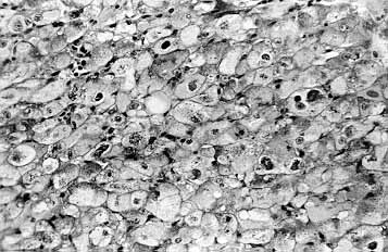
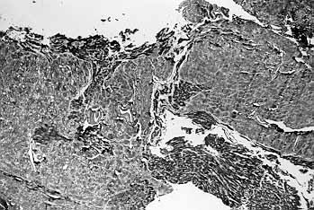 Fig. 2. PTH x 40
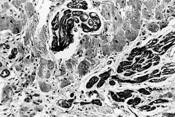 Fig. 3. PTH x 200