
 Comunciación libre
|
PRIMARY IMMATURE MEDIASTINAL TERATOMA IN A DEAD NEWBORN. CASE REPORT Clóvis Klock*, Ivan Tadeu Rebouças* |
|
|
BACKGROUND
Primary immature mediastinal teratomas pose a diagnostic challenge, as they are extremely rare whereas can be cured if promptly diagnosed. The authors report a case of immature mediastinal teratoma in a dead newborn
METHOD AND RESULTS
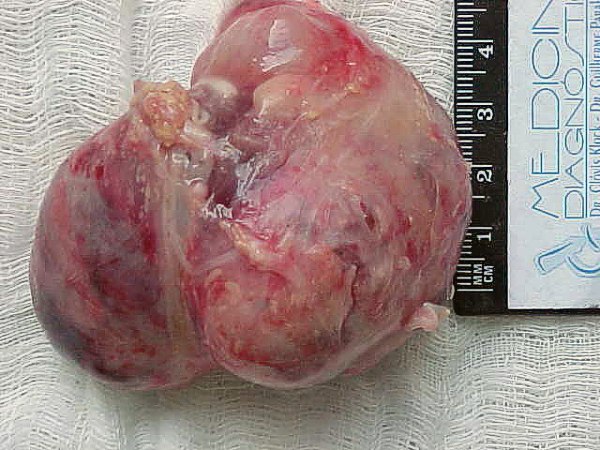
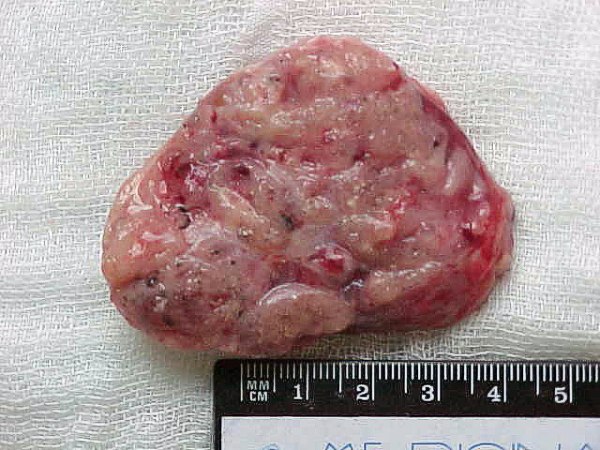
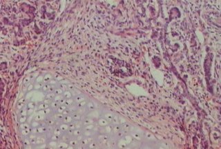
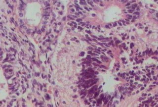
A 36 years old pregnant woman was admitted at 32- weeks gestation and had a cesarean section with an outcome of a dead newborn. A previous ultrasound examination showed a living fetus with generalized edema. A necropsy was performed on the newborn according to the Perinatal Autopsy Manual (AFIP- 1983). Received was a dead fetus of 2780,0 g of weight, measuring 43,0 cm of length. The inspection of the thoracic cavity showed a 88,0 g, 7,5 x 6,0 x 3,5 cm, bosselated, pink - tan tumor mass in thymic topography, displacing the thoracic organs. All tissues were immersion fixed in formalin, routinely processed, embedded in paraffin and were sectioned and stained with hematoxylin and eosin. Histological samples showed mature epithelial and mesenchymal tissues and also mesenchymal and immature neuroepithelium. A diagnosis of immature teratoma was made
CONCLUSION
Primary immature mediastinal teratomas are very rare, comprising 1% of all mediastinal teratomas. Immediately after the delivery, these tumors may cause life threatening respiratory obstruction and must be promptly diagnosed and treated, as its prognosis is excellent, without recurrence on the follow- up.
|
||
|
|
Primary immature mediastinal teratomas pose a diagnostic challenge, as they are extremely rare whereas can be cured if promptly diagnosed. The authors report a case of immature mediastinal teratoma in a dead newborn
|
|
|
|
A 36 years old pregnant woman was admitted at 32- weeks gestation and had a cesarean section with an outcome of a dead newborn. A previous ultrasound examination showed a living fetus with generalized edema. A necropsy was performed on the newborn according to the Perinatal Autopsy Manual (AFIP- 1983). Received was a dead fetus of 2780,0 g of weight, measuring 43,0 cm of length. The inspection of the thoracic cavity showed a 88,0 g, 7,5 x 6,0 x 3,5 cm, bosselated, pink - tan tumor mass in thymic topography, displacing the thoracic organs. All tissues were immersion fixed in formalin, routinely processed, embedded in paraffin and were sectioned and stained with hematoxylin and eosin. Histological samples showed mature epithelial and mesenchymal tissues and also mesenchymal and immature neuroepithelium. A diagnosis of immature teratoma was made
|
|
|
|
Primary germ cell tumors of the mediastinum in newborns are unusual neoplasms with histopathologic features that are similar to those of germ cell tumors in the gonads. In this location, all types of germ cell tumors were described: seminomatous, non-seminomatous, teratomas, nonteratomatous germ cell tumors (yolk sac tumors, embryonal carcinomas, and choriocarcinomas) and combined germ cell tumors without teratomatous components (1) From those types, the most common were mediastinal teratomas. Mediastinal teratomas are uncommon in infants and children, constituting 7% to 10% of all teratomas in this age group (2,3). The overwhelming majority of patients are male (1) In newborns, immature teratomas are rare and constitute less than 1% of all mediastinal teratomas. Other uncommon sites where immature teratomas are reported in early infancy include brain(4), neck(5), pharynx(6), stomach(7) and mesentery(8) Review of literature revealed that respiratory distress is the most common presentation of immature teratoma of mediastinum in newborn (3,4,9). Chest X-ray often demonstrated a mediastinal mass, some of which are calcified, but a definite diagnosis of teratoma is established only after microscopic examination (3). Immature teratoma is characterized by the presence of elements that resemble embryonic tissues, including neuroglial or neuro-epithelial components that may coexist along with mature tissues. In most instances, immature teratomas occurring in the fetus and newborn are associated with a favorable prognosis (2,9,10). The behavior of immature teratoma in adolescents and adults is less predictable and may be associated with poor clinical outcome (9). Dehner cites a 15% overall malignancy rate for mediastinal teratomas diagnosed in the pediatric age group (2). However, other workers concluded that the mediastinal teratomas, both mature and immature, occurring in newborns and infants behave in a benign fashion, if resectable, and are associated with a favorable prognosis as compared to those occurring in adolescents and adults (3,9,11). In our case, the prenatal diagnosis was not made and the fetus died in utero.
|
|
|
|
Primary immature mediastinal teratomas are very rare, comprising 1% of all mediastinal teratomas. Immediately after the delivery, these tumors may cause life threatening respiratory obstruction and must be promptly diagnosed and treated, as its prognosis is excellent, without recurrence on the follow- up.
|
|
|
|
1 - Moran CA, Suster S. Primary germ cell tumors of the mediastinum: I. Analysis of 322 cases with special emphasis on teratomatous lesions and a proposal for histopathologic classification and clinical staging. Cancer. 1997 15;80(4):681-90. 2 - Dehner LP. Gonadal and extragonadal germ cell neoplasia in childhood. Human Pathol 1983; 14: 493-511. 3 - Lakhoo K, Boyle M, Drake DP. Mediastinal teratomas: Review of 15 pediatric cases. J Pediatr Surg 1993; 28: 1161-1164. 4 - Pinto V, Meo F, Loiudice L, D’ Addario V. Prenatal sonographic imaging of an immature intracranial teratoma. Fetal Diagn Ther 1999; 14: 220-222. 5 - Dunn CJ, Nguyen DL, Leonard JC. Ultrasound diagnosis of immature cervical teratoma: A case report. Am J Perinatol 1992; 9: 445- 447. 6 - Mcmohan MJ, Chescheir NC, Kuller JA, Wells SR, Wright LN, Nakayama DK. Peri-natal management of a lingual teratoma. Obstet Gynecol 1996; 87: 848-851. 7 - Sarin YK, Agarwal LD, Jhamaria VN, Goyal RB, Sharma R, Shekhawat NS. Immature gastric teratoma. Indian J Pediatr 1997; 64: 896-898. 8 - Marcolongo A, Divirgilio G, Bettili G, Savero CF, Fasoli L, Marradi P, et al. Immature mesenteric teratoma in a male newborn infant: Prenatal ultrasonographic diagnosis and surgi-cal treatment. Prenat Diagn 1997; 17: 686-688 9 - Carter D, Bibro MC, Touloukian RJ. Benign clinical behavior of immature mediastinal teratomas in infancy and childhood; Report of two cases and review of the literature. Cancer 1982; 49: 398-402. 10 - Isaacs H Hr. Perinatal (Congenital and neonatal) neoplasms: A report of 110 cases. Pediatr Pathol 1985; 3: 165-216.
|
|
|
|
- Oscar Marin (16/10/2005 0:53:26)
- Clóvis Klock (17/10/2005 12:26:07)
|
|
|
|
|
Web mantenido y actualizado por el Servicio de informática uclm. Modificado: 16/06/2015 15:10:50
 fiogf49gjkf0d">
 fiogf49gjkf0d">
 fiogf49gjkf0d">
 fiogf49gjkf0d">
 fiogf49gjkf0d">
 fiogf49gjkf0d