
 Comunciación libre
|
RHINO- CEREBRAL ZYGOMYCOSIS IN A DIABETES MELLITUS PATIENT. CASE REPORT Clóvis Klock*, Ivan Tadeu Rebouças**, Ane Cristine Zenella Monteiro*** |
|
|
BACKGROUND
Zygomycosis is a rare infection, usually affecting patients with diabetes mellitus and neoplasia. The fungi usually do not grow in culture media and the diagnosis depends upon finding the fungi in the anatomopathological samples.
CASE: The case is of a 61 years old adult male with diabetes mellitus type 2
METHODS: The samples were routinely processed and stained with hematoxylin and eosin, PAS, silver staining. A culture of the material was also performed.
RESULTS: Anatomopathological examination – first surgery (28/12/2001): Received were small fragments labeled as from right and left nasal fossae. The histological examination showed respiratory mucosa with edema, vascular congestion, hemorrhage, vascular proliferation, areas of necrosis and purulent exudate. The PAS and silver staining showed hyphae with cytoplasm, amphophilic, non- septated, broad, some short, other long, cenocytic. Some were located within the necrotic material, some around the lumen of the vessels, some invading the vessels themselves. Anatomopathological examination – second surgery (31/01/2002). Received were small fragments of bone and soft tissues from the following sites: right maxillary bone, nasal bone, left and right frontal bones, ethmoid bone, vomer, middle and left inferior nasal conchae and right parotid duct. The anatomopathological samples showed the same findings of the first sampling but also showed invasion of the bone tissues by the fungi
COMMENTS: The patient was treated with amphotericin B. He is been followed up in outpatient basis with good response. The zygomycosis is an extremely rare infection with a very high mortality rate. Its early diagnosis associated with surgical debridement and antifungal treatment were essential to the succesful outcome of this case
|
||
|
|
Zygomycosis is a group of opportunistic infections caused by species of zygomycetes in the order Mucorales. The agents of zygomycosis (or mucormycosis) normally reside in the environment. The abundant airborne sporangiospores may be inhaled or contaminate wounds or burns of patients with predisposing conditions, such as diabetes or immunosuppression, or trauma, and cause a spectrum of diseases. Zygomycosis results from the invasion of the major blood vessels by the fungus, which grows in their walls and lumen causing thromboembolism, ischemia, and necrosis of the tissues. The infection is usually acute and rapidly progressive; however, chronic forms and colonization of some the sinuses and other anatomic sites have been reported .
|
|
|
|
CASE: The case is of a 61 years old adult male with diabetes mellitus type 2. The samples were routinely processed and stained with hematoxylin and eosin, PAS, silver staining. A culture of the material was also performed. Anatomopathological examination first surgery (28/12/2001): Received were small fragments labeled as from right and left nasal fossae. The histological examination showed respiratory mucosa with edema, vascular congestion, hemorrhage, vascular proliferation, areas of necrosis and purulent exudate. The PAS and silver staining showed hyphae with cytoplasm, amphophilic, non- septated, broad, some short, other long, cenocytic. Some were located within the necrotic material, some around the lumen of the vessels, some invading the vessels themselves. Anatomopathological examination second surgery (31/01/2002). Received were small fragments of bone and soft tissues from the following sites: right maxillary bone, nasal bone, left and right frontal bones, ethmoid bone, vomer, middle and left inferior nasal conchae and right parotid duct. The anatomopathological samples showed the same findings of the first sampling but also showed invasion of the bone tissues by the fungi
|
|
|
|
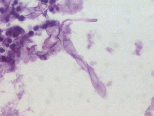
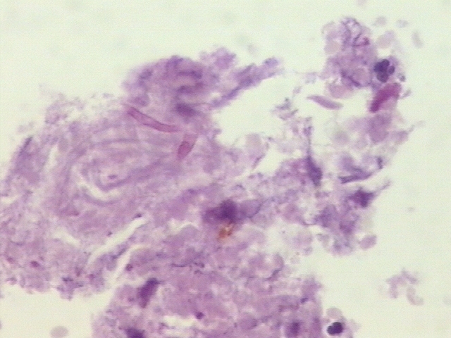
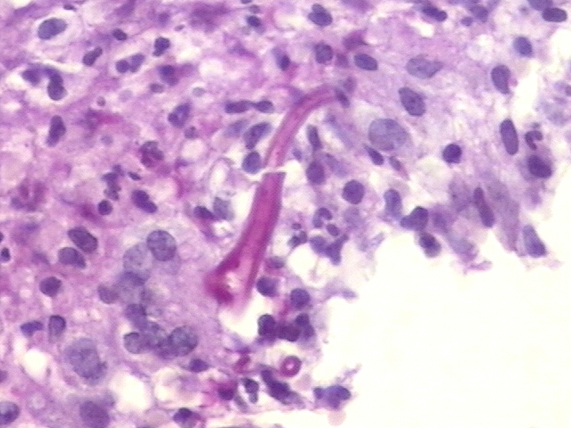
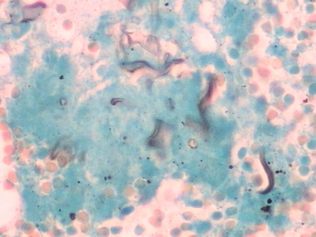
The PAS and silver staining showed hyphae with cytoplasm, amphophilic, non- septated, broad, some short, other long, cenocytic. Some were located within the necrotic material, some around the lumen of the vessels, some invading the vessels themselves
|
|
|
|
Mucormycosis is an acute and often fatal infection caused by a fungus of the Mucorales order of the Zygomycetes class. In the majority of cases, it is associated with an underlying disorder, such as diabetes mellitus with ketoacidosis, or with immunocompromising factors, but it may appear in healthy people, although rarely. Early diagnosis and treatment are critical to prevent an otherwise fatal outcome. Early diagnosis is critical in the prevention of intracranial extension of the infection, which is the cause of death in 80% of cases. Therefore, a high index of clinical suspicion is essential in immunocompromised or diabetic patients with acute sinus infection. Identification of a fungal organism on histopathology is the most specific element for diagnosis. A team approach to management is recommended for early surgical debridement, correction of diabetic ketoacidosis, and systemic antifungal agents. Timely medical-surgical treatment proves extremely important for prognosis.
|
|
|
|
The patient was treated with amphotericin B. He is been followed up in outpatient basis with good response. The zygomycosis is an extremely rare infection with a very high mortality rate. Its early diagnosis associated with surgical debridement and antifungal treatment were essential to the succesful outcome of this case
|
|
|
|
1 - Safar A, Marsan J, et al. Early identification of rhinocerebral mucormycosis. J Otolaryngol. 2005;34(3):166-71.
2 - Basan C III, Rinoldi MG, Rauch RR, Jinkins JR. Fungal infections of the brain. Neuroimaging Clin N Am 1991;1:5758
3 - Davis RL, Robertson DM. Textbook of Neuropathology. 2nd ed. Baltimore: Williams & Wilkins, 1991:761763
4 - Del Rio Perez , Santin Cerezales M, et al. Mucormycosis: a classical infection with a high mortality rate. Report of 5 cases. Rev Clin Esp. 2001;201(4):184-7
5 - Hyvernat H, de Swardt P, et al.Rhino-cerebral mucormycosis complicated by intracerebral hemorrhage with favorable outcome.Presse Med. 2000 Oct 28;29(32):1762.
|
|
|
|
- Emilio Mayayo Artal (03/10/2005 0:29:52)
- Mirta Garcia Jardon (13/10/2005 8:44:35)
- JORGE SAINZ BALLESTEROS (23/10/2005 16:29:23)
- Clóvis Klock (24/10/2005 20:19:09)
- Mirta Garcia Jardon (31/10/2005 17:03:59)
|
|
|
|
|
 fiogf49gjkf0d">
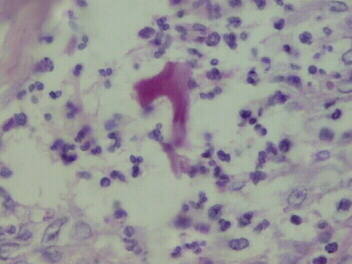 fiogf49gjkf0dHyphae with cytoplasm, amphophilic, non- septated - PAS stain">
.JPG) fiogf49gjkf0d">
fiogf49gjkf0d">
 fiogf49gjkf0d">
 fiogf49gjkf0d">
 fiogf49gjkf0d">
 fiogf49gjkf0d">
 fiogf49gjkf0d">
 fiogf49gjkf0d">
Web mantenido y actualizado por el Servicio de informática uclm. Modificado: 16/06/2015 15:10:50