
 Comunciación libre
|
Roberto Silva Aguiar* |
|||||||||||||||||||||||||||||||||||||||||||||||||||||||||||||||||||||||||
|
Objetivos: Describir y validar una experiencia de 10 años con citologías aspirativas con aguja fina (CAAF) en niños y adolescentes. Diseño del estudio: La CAAF fue realizada principalmente como citopunción por un patólogo. Los archivos de citopatología del Hospital Pediátrico William Soler fueron revisados y se seleccionó los registros de los pacientes menores de 21 años. Se creó una base de datos con la información usando el programa Excel. Se revisó también los datos clínicos e histopatológicos. La base de datos fue analizada y se realizó cálculos, gráficos y tablas. Resultados: 3274 CAAF fueron realizadas con predominio del sexo masculino (55%) y de niños menores de 7 años. Los Ganglios linfáticos, Partes blandas , Tiroides y Mama fueron los sitios más frecuentemente puncionados. Hubo 80.9% de diagnósticos benignos, 6% malignos, 2.4% sospechosos de malignidad y 10.7% insuficientes para diagnóstico. 630 citologías fueron correlacionadas: 69 fueron insuficientes, 321 verdaderos negativos, 184 verdaderos positivos, 19 falsos negativos y 37 falsos positivos. La sensibilidad, especificidad, precisión, valores predictivos positivo y negativo y prevalencia fueron 90.6%, 89.6%, 90%, 83.2% 94.4%, y 36.1% respectivamente. 4 complicaciones de las punciones en órganos profundos fueron observadas y el índice de complicaciones fue de 0.01%. Conclusión: La CAAF es una excelente técnica diagnóstica en niños y adolescentes como en adultos. (Objectives: To describe and to validate a 10-year experience in pediatric fine needle aspiration citology (FNAC). Study design: FNAC was done mainly in a non aspirating form by a pathologist. Archives of cytopathology at William Soler Pediatric Hospital were reviewed and patients punctured younger than 21 years were selected. A data base was created with patients information using Excel program. Histopathology and clinical data were also reviewed. Data base was analyzed, figures, graphics and tables were done as needed. Results: 3274 fine needle cytologies were done. There was a predominance of male patients (55%) and children younger than seven years. Lymph nodes, soft tissue, thyroid, and breast were the most frequently punctured sites. There were 80,9% benign diagnosis, 6% malignant, 2,4% suspicious for malignancy and 10,7% insufficient. 630 cytologies were correlated: 69 were insufficient, 321 true negative, 184 true positive, 19 false negative, and 37 false positive. Sensibility, specificity, accuracy, positive and negative predictive values and prevalence were 90.6%, 89.6%, 90%, 83.2%, 94.4%, and 36.1%. Four deep organ puncture complications were observed and complication rate was 0,1%. Conclusion: FNAC is an excellent diagnostic technique in pediatric age as in adulthood.)
|
||||||||||||||||||||||||||||||||||||||||||||||||||||||||||||||||||||||||||
|
|
Fine needle aspiration cytology (FNAC) in adult age is a well recognized diagnostic technique with accepted value in management of tumoral and pseudotumoral processes 1-3. Nevertheless its use in children and teenagers is still underused 4-6. Some reports about FNAC employment in the early years of life advocate its value in a very similar way as in adulthood 7-10 but many pediatricians reject it arguing poor child cooperation during the puncture, the need of sedation, the inability to obtain an adecuate sample and the intrinsic diagnostic difficulties of small round blue cell tumors of infancy. It is our purpose to demonstrate our experience with the use of FNAC in a major children hospital and prove its statistical value. We havent found any previus pediatric report with such a diversity of sites punctured.
|
|||||||||||||||||||||||||||||||||||||||||||||||||||||||||||||||||||||||||
|
|
Previous approbation of this research by the local institutional review board, archives of cytopathology at William Soler Pediatric Hospital were reviewed from January 1992 to September 2002. An Excel data base was made with data from all punctured patients younger than 21 years. Later, histopathology archives were reviewed to correlate cytology and histology. Evolutive clinical data (1-118 moths) of punctured cases without biopsy were also correlated. Statistical results were obtained analyzing the data base. Figures, tables and graphics were done as needed 11. The punctures without anesthesia were mainly done by a pathologist with experience and special interest in this area according to established standards 1,3,12. The cytologic samples obtained by puncture were expelled and smeared onto glass slides and stained with May Grünwald Giemsa or Hematoxylin-Eosin. Diagnosis was done knowing clinical data and pertinent laboratory and image information when available. Thoracic and mediastinal punctures under thoracoscopic guide were performed by surgeons under anesthesia. Deep abdominal tumors were punctured with or without real time ultrasonography but without guide, sedation or anesthesia. Cytology diagnoses were classified as benign (neoplastic or not), malignant (with or without histogenetic approach), suspected of malignancy (intermediate diagnostic category not enough to make a malignant diagnosis of certitude) and insufficient.
|
|||||||||||||||||||||||||||||||||||||||||||||||||||||||||||||||||||||||||
|
|
Distribution of punctures by organ or site
|
|||||||||||||||||||||||||||||||||||||||||||||||||||||||||||||||||||||||||
|
|
Pediatric FNAC is nowadays an increasingly used technique. Literature review shows that pure pediatric series are rare, some of them covering up to 300 patients of different ages. It has been our objective to analyze and present a first report with 3274 FNAC done in 22 sites or organs.
|
|||||||||||||||||||||||||||||||||||||||||||||||||||||||||||||||||||||||||
|
|
In conclusion: FNAC can be used in pediatrics in a similar way as it is employed in adult age.
|
|||||||||||||||||||||||||||||||||||||||||||||||||||||||||||||||||||||||||
|
|
1 -Buley ID, Roskell DE: Fine-needle aspiration cytology in tumour diagnosis: uses and limitations. Clin Oncol (R Coll Radiol) 2000;12(3):166-71 2 -Jaffer S, Zakowski M: Fine-needle aspiration biopsy of axillary lymph nodes. Diagn Cytopathol 2002 ;26(2):69-7 3 -Amedee RG, Dhurandhar NR: Fine-needle aspiration biopsy. Laryngoscope 2001;111(9):1551-7 4 -Layfield LJ, Reichman A: Fine needle aspiration cytology: utilization in pediatric pathology. Dis Markers 1990;8(6):301-15 5 -Howell LP: Chanching role of Fine needle aspiration in the evaluation of pediatric masses. Diagn Citopathol 2001;24:65-70 6 -Maramo I, Soscia E, Salvatore M: Abdominal mass in childhood: Characterization with fine needle percutaneous biopsy guided with computarized tomography. Radiol Med (Torino) 1999;97(4):251-5 7 -Valkov I, Bojikin B: Fine-needle aspiration biopsy of abdominal and retroperitoneal tumors in infants and children. Diagn Cytopathol 1987;3(2):129-33. 8 -Wakely PE Jr, Kardos TF, Frable WJ: Application of fine needle aspiration biopsy to pediatrics. Hum Pathol 1988;19(12):1383-6. 9 -Silverman JF, Gurley AM, Holbrook CT, Joshi VV: Pediatric fine-needle aspiration biopsy. Am J Clin Pathol. 1991;95(5):653-9 10 -Rajwanshi A, Rao KL, Marwaha RK, Nijhawan VS, Gupta SK.: Role of fine-needle aspiration cytology in childhood malignancies. Diagn Cytopathol. 1989;5(4):378-82. 11 -Rabb S: Diagnostic accuracy in cytopathology. Diagn Cytopathol 1994 10(1):68-75 12 -National Committee for clinical laboratory standards. Fine Needle Aspiration Biopsy (FNAB) techniques: Approved guidelines - Villanova, PA: NCCLS, 1997.NCCLS document GP20A 13 -Orford JE, Gollow I, Brennan B, Hallam L: Fine needle aspiration biopsy in children. Aust N Z J Surg 1997;67(11):785-8 14 - Gamba PE, Messineo A, Antoniello LM, Boccato P, Blandamura S, Cecchetto G, Dalla'Igna P, Guglielmi M: A simple exam to screen superficial masses: Fine needle aspiration. Med Pediatr Oncol 1995;26:97-99 15 -Almeida MM, Mendonca ME, Sousinha M, Martins AAG, de Sousa JV, Soares J: Application of aspiration cytology in the diagnosis of tumor lesions in children. Acta Med Port 1991;4(2):71-5 16 -Millar AJ, Sinclair-Smith G, Rode H, Hartley P, Karabus C, Cywes S: Fine needle cytology of solid tumors: Method, diagnostic accuracy ad role in management. J Pediatr Surg 1990;25(10):1088-91 17 -Smith MB, Katz R, Black CT, Cangir A, Andrassy RJ: A rational approach to the use of fine-needle aspiration biopsy in the evaluation of primary and recurrent neoplasms in children. J Pediatr Surg. 1993;28(10):1245-7 18 -Eisenhut CC, King DE, Nelson WA, Olson LC, Wall RW, Glant MD: Fine needle biopsy of pediatric lesions: A tree year study in an outpatientbiopsy clinic. Diagn Cytopathol 1996;14(1):43-50 19 -Nesbit ME: Clinical assesment of differential diagnosis of the child with suspected cancer. In: Pizzo PA, Poplack DG. Principles and practice of pediatric oncology, 2nd edition, Philadelphia, JB Lippincott Company 1993,109-110 20 -Verdeguer A, Castel V, Torres V, Olague R, Ferris J, Esquembre C, Vallcanera A, Muro MD: Fine-needle aspiration biopsy in children: experience in 70 cases. Med Pediatr Oncol. 1988;16(2):98-100 21 -Obers VJ, Phillips JI: Fine needle aspiration of pediatric abdominal masses. Cytologic and electron microscopic diagnosis. Acta Cytol. 1991;35(2):165-70. 22 -Tayler Sr, Nunez C: Fine neddle aspitarion biopsy in a pediatric population. Report on 64 consecutive cases. Cancer 1984;54:1449-53 23 -Diament MJ, Stanley P, Taylor S: Percutaneous fine needle biopsy in pediatrics.Pediatr Radiol 1985;15(6):409-11 24 -Cohen MB, Bottles K, Ablin AR, Miller TR: The use of fine-needle aspiration biopsy in children. West J Med. 1989 Jun;150(6):665-667 25 -Schaller RT Jr, Schaller JF, Buschmann C, Kiviat N: The usefulness of percutaneous fine-needle aspiration biopsy in infants and children. J Pediatr Surg 1983;18(4):398-405 26 -Jereb B, Us-Krasovec M, Jereb M: Thin needle biopsy of solid tumors in children. Med Pediatr Oncol. 1978;4(3):213-20 27 -Layfield LJ, GlasgowB, Ostrzega N, Reynolds CP: Fine needle aspiration cytology and the diagnosis of neoplasm in the pediatric age group. Diagn Cytopathol 1991;7:451-461 28 -Gorczyca W, Bedner E, Juszkiewicz P, Chosia M: Aspiration cytology in the diagnosis of malignant tumors in children. Am J Pediatr Hematol Oncol 1992;14(2):129-34; discussion 134-5 29 -Howell LP, Russell LA, Howard PH, Teplitz RL: The cytology of pediatric masses: a differential diagnostic approach. Diagn Cytopathol 1992;8(2):107-15 30 -McGahey BE, Moriarty AT, Nelson WA, Hull MT: Fine-needle aspiration biopsy of small round blue cell tumors of childhood. Cancer 1992;15;69(4):1067-73 31 -Geisinger LR, Silverman JF, Wakely PE. Pediatric Cytopathology. William Johnston MD series editor, Duke University ASCP Theory and Practice of Cytopathology Vol 4 Chicago: American Society of Clinical Pathologist Press; 1994,1-9
|
|||||||||||||||||||||||||||||||||||||||||||||||||||||||||||||||||||||||||
|
|
- Julia Cecilia Cruz Mojarrieta (02/10/2005 15:47:10)
- Santiago Quintero (10/10/2005 22:51:42)
- JORGE SAINZ BALLESTEROS (20/10/2005 12:26:19)
- Juan Carlos Perez-Cardenas (25/10/2005 3:47:52)
- Manuel Diaz-Marta Puentes (14/10/2011 5:29:52)
- Manuel Diaz-Marta Puentes (14/10/2011 5:29:53)
- Manuel Diaz-Marta Puentes (14/10/2011 5:29:54)
- Manuel Diaz-Marta Puentes (18/02/2012 6:38:23)
- Manuel Diaz-Marta Puentes (18/02/2012 6:38:23)
- Manuel Diaz-Marta Puentes (18/02/2012 6:38:24)
|
|||||||||||||||||||||||||||||||||||||||||||||||||||||||||||||||||||||||||
|
|
|
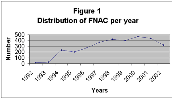 fiogf49gjkf0dFigure 1.">
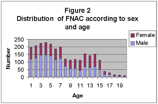 fiogf49gjkf0dFigure 2.">
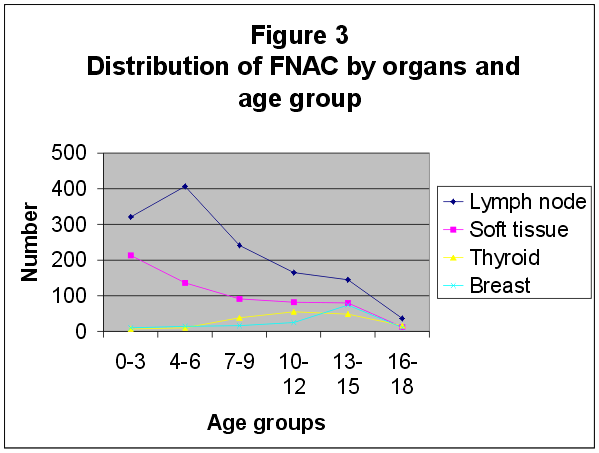 fiogf49gjkf0dFigure 3.">
Web mantenido y actualizado por el Servicio de informática uclm. Modificado: 16/06/2015 15:10:50