

|
Quiste postraumático del bazo: Reporte de un caso y revisión de la literatura. Walter Marcial Martínez Rodríguez*, María de los Ángeles Miló Anillo*, Aldo Sisto Díaz**, Magalis Rodríguez***, Raul Rua Martínez*, Mayda Martínez Rodríguez****, Raul Rua González****, Ana Gloria Pérez Reyes***** |
||
|
Los quistes esplénicos son lesiones raras. Ellos se dividen principalmente en primarios o genuinos y secundarios o falsos, de acuerdo con su etiología y fisiopatología. Los quistes primarios tienen un revestimiento epitelial que puede ser causado por o eventos primarios o infestaciones parasitarias (Equinococos). Los quistes secundarios no tienen revestimiento epitelial y pueden ser: hemorrágicos, serosos, inflamatorios, o degenerativos. Es importante para el cirujano poder evaluar bien cada caso individual para poder decidir sobre el tratamiento más aconsejable, teniendo en cuenta: las características del quiste, el debut, y la edad del paciente; para evitar posibles complicaciones. Reportamos un caso de quiste postraumático del bazo tratado con esplenectomía total; el cual evolucionó satisfactoriamente
|
|||
|
|
Los quistes esplénicos son lesiones raras. Ellos se dividen principalmente en primarios o genuinos y secundarios o falsos, de acuerdo con su etiología y fisiopatología. Los quistes primarios tienen un revestimiento epitelial que puede ser causado por o eventos primarios o infestaciones parasitarias (Equinococos). Los quistes secundarios no tienen revestimiento epitelial y pueden ser: hemorrágicos, serosos, inflamatorios, o degenerativos. Es importante para el cirujano poder evaluar bien cada caso individual para poder decidir sobre el tratamiento más aconsejable, teniendo en cuenta: las características del quiste, el debut, y la edad del paciente; para evitar posibles complicaciones. Reportamos un caso de quiste postraumático del bazo tratado con esplenectomía total; el cual evolucionó satisfactoriamente (1)
|
||
|
|
Caso Clínico, paciente de 53 años, del sexo masculino, de la raza blanca, que comenzó a sentir dolor en hipocondrio izquierdo, al examen físico del abdomen se encontró un bazo palpable y al realizarse un US de hemiabdomen superior se encontró una lesión nodular ecogénica en bazo. El cirujano lo valora y decide hacerle una esplenectomía parcial. En el Departamento de Anatomía Patológica se recibe un segmentode bazo que pesa 160 gms, superficie lisa violácea, presenta un área focal renitente que protruye y confiere a la cápsula esplénica una coloración amarillenta focal en su cara diafragmática (figura1) al corte se encontró una lesión quística de 5 x 3.5 cm ovalada y totalmente calcificada (figura 2 y 3), unilocular, de contenido líquido, seroso transparente, con detritus celulares; en cantidad de 5cc; la pared midió 1mm. El contenido líquido fue centrifugado y examinado minuciosamente al microscopio no encontrándose restos parasitarios de ningún tipo; solo encontramos detritus celulares, histiocitos fagocíticos, muchos de ellos cargados de hemosiderina. La pared no presentó ningún revestimiento epitelial pudiendo observarse, después de la descalcificación, que estaba constituida por tejido de granulación con fibrosis densa periférica que se había colagenizado.
|
||
|
|
En Cuba no tenemos equinococosis, los casos que hemos tenido han sido casos importados, por esta razón los quistes parasitarios son muy raros aunque siempre que nos encontremos con un quiste esplénico debemos descartar la posibilidad de una equinococosis, después de descartarla y quedarnos con un quiste no parasitario tenemos que ver si es primario (verdadero) o secundario (falso). (2)
|
||
|
|
1-Labruzzo C, Haritopoulos KN, El Tayar AR, Hakin NS. Posttraumatic cyst of the spleen; a case report and review of the literature. Int Surg. 2002 Jul-Sep;87(3):152-6. 2-González I, Díaz M, Ángel F y González O. Infección por Ecchinococcus granulosus (quiste hidatídico). Reporte de un caso. Rev Cub Med Trop 2001; 53(3): 217-21. 3- Morgenstern, L. Non Parasitic Splenic Cysts: Pathogenesis, Classification, and Treatment. J Am Coll Surg 2002;194(3):306-14
|
||
|
|
- Mario Miguel Morales Wong (08/10/2005 20:20:15)
- Mario Michel Gómez Hernández (08/10/2005 20:20:35)
|
||
|
|
|
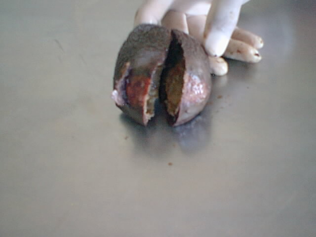 fiogf49gjkf0dBazo parcialmente cortado donde se puede apreciar que el quiste traumático hacía prominencia en su cara diafragmática y le confería a la cápsula una coloración amarillenta focal">
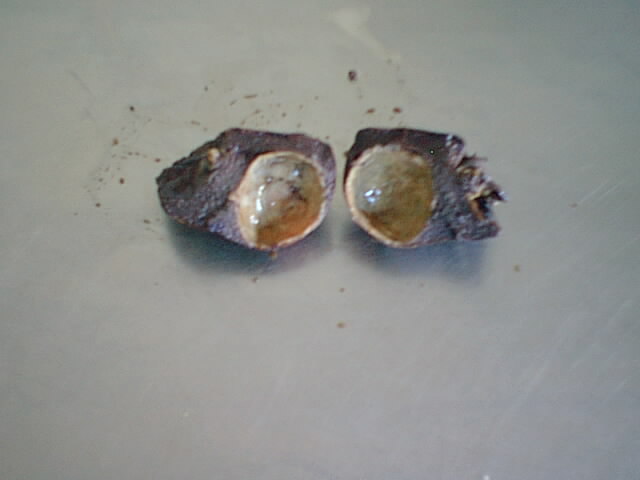 fiogf49gjkf0dEn la superficie de sección (de lejos) se puede apreciar como aparece empotrado, en la pulpa esplénica, un quiste, de pared delgada totalmente calcificada, que recuerda un cascarón de huevo">
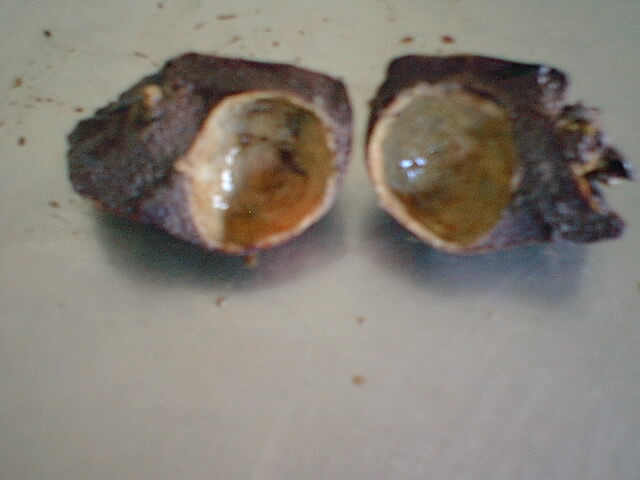 fiogf49gjkf0dEn un acercamiento vemos el "cascarón", mucho mejor">
Web mantenido y actualizado por el Servicio de informática uclm. Modificado: 16/06/2015 15:10:50