|
Virtual Slide Congress. Case # 05
Alexandra Gené*, C Gómez*, A Forteza*, F Terrasa*, M Company*, B Tomás**, M Couce*, Ramón Canet*
* Servicio de Anatomía Patológica. Hospital Universitario Son Dureta. Palma de Mallorca
ESPAÑA
** Servicio de Otorrinolaringología. Hospital Universitario Son Dureta. Palma de Mallorca
ESPAÑA
|
|
Resumen
fiogf49gjkf0d A sphenoidal-sinus lesion in a 55 year-old man.
|
|
Case Report
fiogf49gjkf0d We present a sphenoidal-sinus lesion in a 55 year-old man.
|
|
DIAGNOSIS
fiogf49gjkf0d SPHENOIDAL-SINUS HEPATOCARCINOMA METASTASIS
|
|
Pathology
fiogf49gjkf0d Pathologic findings:
We received two tissue samples that measured 7 and 9 mm.
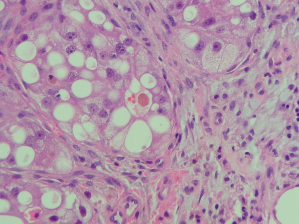
Histologically, broad necro-haemorragic areas with tumoral appearance could be seen.
The periphery of those areas showed poligonal cells with wide cytoplasms, micro and macro esteatotic vacuolization. Some had big vesiculous nuclei with prominent nucleoli that resembled hepatocites. In some areas they adopted a nodular pattern, surrounded by fibrosis with a prominent vascular net. Small bile clusters could also be identified.
.jpg) fiogf49gjkf0d"> fiogf49gjkf0d">
Figura 1 - fiogf49gjkf0d
 fiogf49gjkf0d"> fiogf49gjkf0d">
Figure 2 - fiogf49gjkf0d
|
|
Discusión
|
|
Conclusiones
|
|
Bibliografía
1.- Aung TH, Po YC, Wong WK. Hepatocellular carcinoma with metastasis to the skull base, pituitary gland, sphenoid sinus, and cavernous sinus. Hong Kong Med J. 2002 Feb;8(1):48-51.
2.- Kleinjung T, Held P. Metastasis in the frontal skull base from hepatocellular carcinoma. HNO. 2001 Feb;49(2):126-9.
3.- Sim RS, Tan HK. A case of metastatic hepatocellular carcinoma of the sphenoid sinus. J Laryngol Otol. 1994 Jun;108(6):503-4.
4.- Waxman JS, Seife B, Waxman M. Hepatocellular carcinoma presenting as sphenoid sinus metastasis. Mt Sinai J Med. 1985 Mar;52(3):221-4.
|
|