|
Virtual Slide Congress. Case # 01
Dr. Hugo Góngora Jara*, Dr. Pedro de la Cuesta*, Dr. Aarón Arnaldo Kaen*, Dr. Yoshihiro Matsuno**, Dr. Tadakazu Shimoda**
* Hospital Regional Dr. E Vera Barros. Facultad de Medicina - Instituto Universitario de Ciencias de la Salud Fundación H.A. Barceló. La Rioja. ARGENTINA
** National Cancer Center Hospital, Tokyo. JAPON
|
|
Title
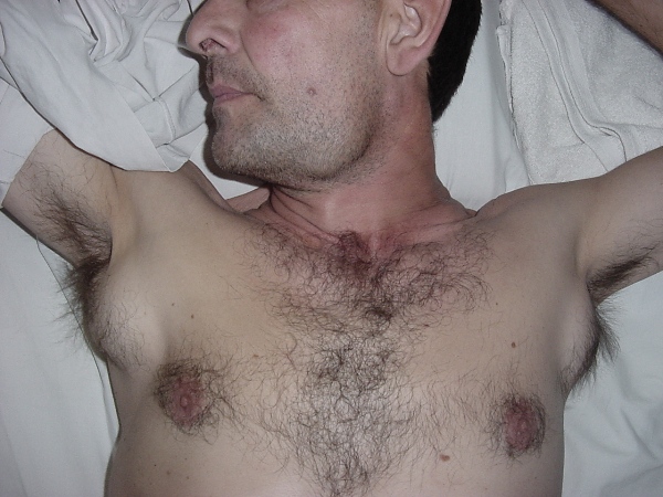
fiogf49gjkf0d Male 49 y.o. showing multiple adenopathy of 1 month of evolution
|
|
Case Report
fiogf49gjkf0d
Male of 49 years that was hospitalized on Medical Clinic Service, showing multiple adenopathy of 1 month of evolution, pain, fever and protracted sweats in the last 3 weeks. The patient lost 10 kg of weight in the last two months.
Poliadenophaty in right preauricular localization of 1cm., left retroauricular side of 3x4cm., left submaxillary side of 6x5cm., submentonian of 2 cm., supraclavicular of 5x3cm., in right axilar region of 5x8 cm., in left axilar region of 5x7cm. and below of left crural arcade of 5x6cm. All lymph nodes presented smooth surface, hard-elastic consistency, adhered to deep planes, and painful to the palpation.
Laboratory
Hemogram Hto 28% Hb 10.
Hepatogram Bilirrubine (normal), Alkaline Phospatase 728 (nv:<270u/l),GOT-GPT (normal), LDH 782 (nv:207 414 u/l). The other laboratory findings were normal.
Chest and abdominal T.A.C.
Lobulated hiliar pulmonary right mass. Extensive ganglionar involvement, mediastinal infracarinal and retrocava. Liver without abnormalities.
Neck T.A.C.
Submaxillary left Adenomegaly, adenopathy conglomerate in left jugulo-carotid chain.
 fiogf49gjkf0d"> fiogf49gjkf0d">
- fiogf49gjkf0d
|
|
DIAGNOSIS
fiogf49gjkf0d Dendritic Cell Sarcoma NOS
|
|
Pathology
fiogf49gjkf0d MAIN PATHOLOGIC FINDINGS:
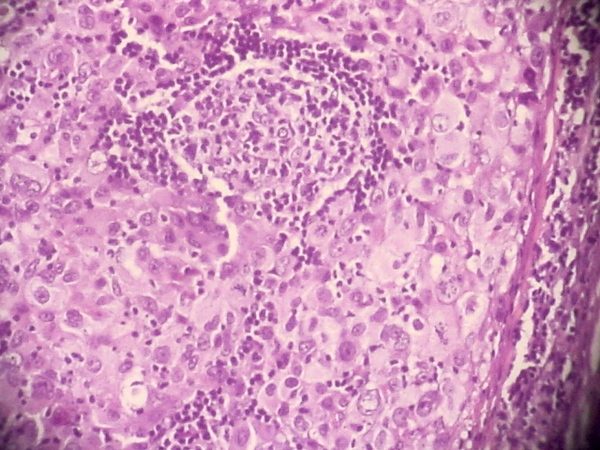
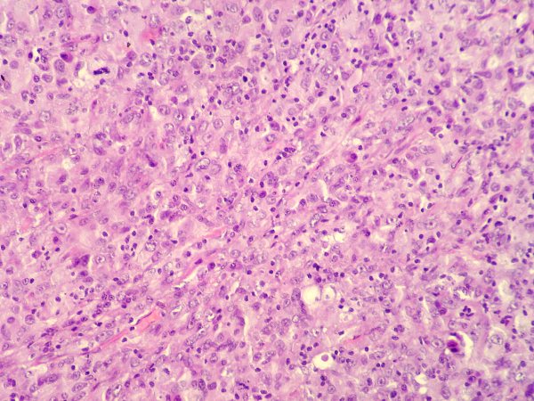
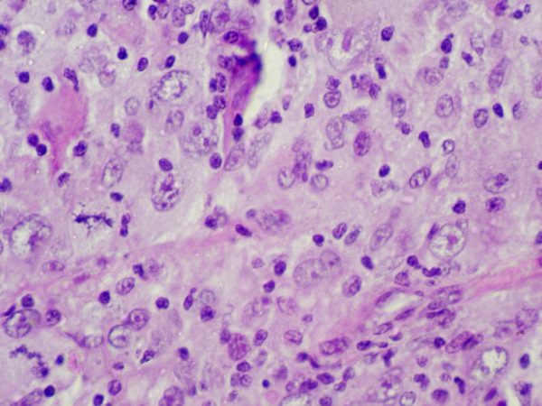
Microscopically, the normal architecture was effaced with diffuse proliferation of large pleomorphic cells arranged in a difusse pattern and rarely and scanty spindle cells with light fascicular distribution. The neoplastic cells had smooth nuclear membranes with round-ovoid to irregularly indented form and small central eosinophilic nucleoli. These cells had moderated to abundant citoplasm, some of theme with slender citoplasmic extensions. Eventually multinucleated cells where found. A constant feature was the presence of variable number of small lymphocites and some plasma cells scattered throughout the tumor. The exceptionally nontumorous area was characterized by reactive germinal center of cortical follicle. Erythrophagocytosis was eventually observed. The mitotic rate was high (30 mitotic figures per 10 high-power fields).
Immunohistochemical studies showed that all of the tumor cells were positive for vimentin, and focal positive for EMA; but uniformly negative for CD45, B and T-cell markers, ALK1, CD30, CD68, Lysozyme, S-100 protein, CD1a, CD21, CD35, Alfa-SMA, Melan-A and AE1-AE3 (CTK Cocktail). In the based on these findings, and the clinic manifestation, the present case was diagnosed as Dendritic Cell Sarcoma not otherwise specified.
 fiogf49gjkf0d"> fiogf49gjkf0d">
Figure 1 - fiogf49gjkf0d
 fiogf49gjkf0d"> fiogf49gjkf0d">
Figure 2 - fiogf49gjkf0d
 fiogf49gjkf0d"> fiogf49gjkf0d">
Figure 3 - fiogf49gjkf0d
|
|
Discusión
|
|
Treatment
fiogf49gjkf0d CHOP protocol.
|
|
Agradecimientos
1.- Al Dr. Carlos Omar Robador, Director General del "Hospital Regional Dr. Enrique Vera Barros" y a la Directora Médica, Dra. Alicia Viale; por facilitar al Servicio de Anatomia Patológica del Hospital la realización de los estudios Inmunohistoquimicos.
To Dr. Carlos Omar Robador, General Director from "Dr. Enrique Vera Barros Regional Hospital" and to Dra. Alicia Viale, Medical Director; because they facilitate the Immunohistochemistry to the Pathology Department.
2.- Al Departamento de Biblioteca de la Fundación H.A. Barceló y por su intermedio al Rector de la Universidad, Prof. Dr. H.A. Barceló, por la obtención de material bibliográfico.
To the Library of "H.A. Barceló Fundation" and to Rector of the University, Prof. Dr. H.A. Barceló, by bibliographical material facilitates.
3.- A JICA(Agencia Cooperativa Internacional del Japón) y a los Profesores Japoneses, por darme la oportunidad de compartir y hacer la rotación en Patología, en la Universidad de Odontología y Medicina de Tokyo y el Centro Nacional del Cáncer de Japón.
To JICA(Japanese International Cooperative Agency) and to Japanesse Professors, for give me the opportunity of share and make trainning in Pathology, in the Dental and Medical University of Tokyo and The National Cancer Center of Japan.
|
|
Bibliografía
Andriko JW, Kaldjian EP, Tsokos M, Abbondanzo SL, Jaffe ES. Reticulum cell neoplasms of lymph nodes: a clinicopathologic study of 11 cases with recognition of a new subtype derived from fibroblastic reticular cells. Am J Surg Pathol. 1998; 22(9):1048-58.
Grogg KL, Lae ME, Kurtin PJ, Macon WR. Clusterin Expression Distinguishes Follicular Dendritic Cell Tumors From Other Dendritic Cell Neoplasms: Report of a Novel Follicular Dendritic Cell Marker and Clinicopathologic Data on 12 Additional Follicular Dendritic Cell Tumors and 6 Additional Interdigitating Dendritic Cell Tumors. Am J Surg Pathol. 2004; 28(8):988-998.
Pileri SA, Grogan TM, Harris NL, Banks P, Campo E, Chan JK, Favera RD, Delsol G, De Wolf-Peeters C, Falini B, Gascoyne RD, GAulard P. Gatter KC, Isaacson PG, Jaffe ES, Kluin P, Knowles DM, Mason DY, Mori S, Muller-Hermelink HK. Tumours of histiocytes and accessory dendritic cells: an immunohistochemical approach to classification from the International Lymphoma Study Group based on 61 cases. Histopathology. 2002; 41(1):1-29.
Vos JA, Abbondanzo SL, Barekman CL, Andriko JW, Miettinen M, Aguilera NS. Histiocytic sarcoma: a study of five cases including the histiocyte marker CD163. Mod Pathol. 2005;18:693-704.
|
|