Congreso Virtual sobre Anatomía Patológica

Virtual Slide Congress. Case # 02

LLUÏSA MORELL QUADRENY*, LAIA BERNET VEGUÉ*, MARIA NAVARRO HERVAS*, RAFAEL CANO MUÑOZ*, XAVIER GISBERT AGUILAR*
* HOSPITAL DE LA RIBERA ALZIRA-VALENCIA ESPAÑA

Resumen

17-year-old male. Dizziness 1 year of evolution
(Varón de 17 años que consulta por síndrome vertiginoso de 1 año de evolución).


Case Report    

fiogf49gjkf0d
17 years-old male complaining of dizziness in the last year. He presented 4-5 episodes of 3-4 days of duration each. Slight subjetive hypoacusia of his right ear. No other personal history of interest.
 
His father suffers from von Hippel Lindau’s disease.
 
ORL, eyes-movement, and neurological physical examinations were normal. Posterior labyrinth examination showed a compensated right vestibular arreflexia.
 
A permeative lesion in the petrous portion of the temporal bone, affecting the vestibular aqueduct was detected in a CT Scan.
 
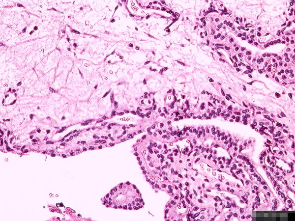
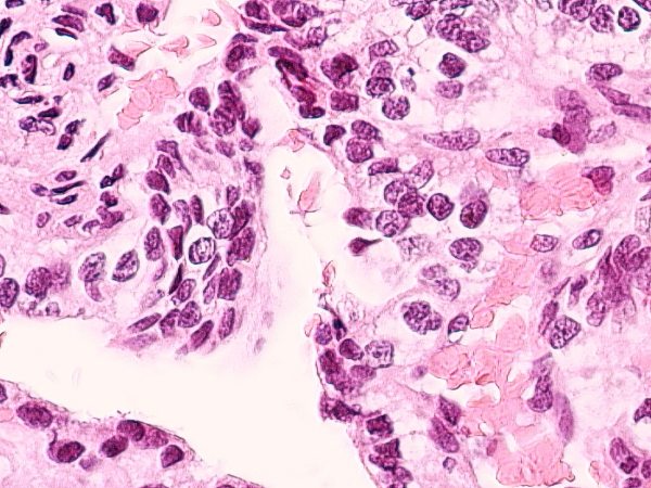
After surgical removal of the tumor, five small hemorrhagic fragments between 0.01 and 0.1 cm were submitted for pathological examination. A separte fibrous 0.5 cm fragment, labelled as durameter, was also received.

 
Varón de 17 años que consulta por síndrome vertiginoso de 1 año de evolución. Refiere  haber presentado 4-5 episodios de 3-4 días de duración. Hipoacusia subjetiva leve de OD. Sin otros antecedentes personales de interés.
 
Padre con enfermedad de von Hippel Lindau.
 
La exploración ORL, del sistema óculo-motor y neurológica son normales. La exploración del laberinto posterior detecta una arreflexia vestibular derecha compensada.
 
Se realiza TAC de peñascos observando en oído derecho una lesión permeativa en la porción pedrosa del hueso temporal, centrada en el acueducto vestibular.
 
Se practica cirugía remitiéndose a anatomía patológica, referenciado como tumor, cinco fragmentos de tamaños comprendidos entre 0,01 y 0,1 cm., de aspecto hemorrágico y, a parte, referenciado como duramadre, un fragmento fibroso de 0,5 cm.


DIAGNOSIS    

fiogf49gjkf0d
Endolymphatic sac tumor (Tumor del saco endolinfático)


Pathology    

fiogf49gjkf0d
Histological study shows, in a dense connective tissue stroma, small papillary and follicular structures containing amorphous eosinophilic material. Those structures are covered by a cubic epithelium, with scarce clear cytoplasm, without any atipia or mitosis.
 
Immunohistochemucal profile of the lesion was:
  • Citokeratin and EMA… Positive
  • Phyroglobulin …………Negative   
 

El estudio microscópico muestra, en un estroma conectivo denso, pequeñas estructuras papilares y foliculares con contenido esosinófilo amorfo.. Ambas se hallan tapizadas por un epitelio cúbico, con escaso citoplasma claro, sin atipia ni mitosis.
El perfil inmunohistoquímico de la lesión es: 
  • Citoqueratina y EMA: Positivas.
  • Tiroglobulina: .............Negativa.


  Figure 1 - <div style=fiogf49gjkf0d">
Figure 1 -

fiogf49gjkf0d


  Figure 2 - <div style=fiogf49gjkf0d">
Figure 2 -

fiogf49gjkf0d


Discusión    

Conclusiones    

Agradecimientos    

Bibliografía    

1.- H Jeffrey Kim, JA Butman, C Brewer, Ch Zalewski, AO Vortmeyer, G Glenn, E H Oldfield, R L Lonser: Tumors of the endolymphatic sac in patients with von Hippel-Lindau disease: implications for their natural history, diagnosis and treatment. J neurosurg 102: 503-512, 2005-06-23

2.- D A LUF, M Simmons, T Malik, R T Ramsden, H Reid, C Path: Endolymphatic sac tumours. The Journal of Laryngology & Otology 116: 398-401, 2002

3.- G Kempermann, HP Neumann, Volk B: Endolymphatic sac tumours. Histopathology 33(1): 2-10, 1998


Web mantenido y actualizado por el Servicio de informática uclm. Modificado: 16/06/2015 15:17:23