|
|
Image cytometry in the gastrointestinal tract precancerous lessions
Dr Javier Azúa Romeo*
,
Dr. Javier Azúa Blanco*
,
Dra. María Peña Romeo Casabona**
,
Dra Mayte Tovar Lázaro*
y
Dr Rafael Uribarrena ^
*Departamento de Anatomía e Histología Humanas.Universidad de Zaragoza
** Profesor de Biofísica. Universidad de Zaragoza
*** Mutua de Accidentes de Zaragoza
^Sº Aparato Digestivo Hospital Miguel Servet. Zaragoza
España
|
|
|
|
|
|
|
|
Resumen
|
|
|
|
DNA quantification has a recognized value as a prognostic factor in certain human neoplasms; nevertheless, there are very few references regarding the use of this technique applied over dysplastic lesions, moreover we haven´t found any report about gastrointestinal tract dysplasias. It is our thought that DNA quantification in digestive cancer has been determinant for the establisment of more adequate therapy, fiting the surgical treatment to the biological behaviour of the tumor and the survival expectation.
The use of this tool for dysplastic lesions may suppose an step forward in the fight against cancer, since it could be performed after the practice of a fiber-endoscopy, thus selecting patients with higher risk of developing malignancy or else classifying tumors as already a truly cancer rather than dysplasia.
|
|
|
|
|
|
|
Introducción
|
|
|
|
DNA quantification has a recognized value as a prognostic factor in certain human neoplasms; nevertheless, there are very few references regarding the use of this technique applied over dysplastic lesions in the gastrointestinal tract 1,2. It is our thought that DNA quantification in digestive cancer has been determinant for the establisment of more adequate therapy, fiting the surgical treatment to the biological behaviour of the tumor and the survival expectation 3,4.
The use of this tool for dysplastic lesions may suppose an step forward in the fight against cancer, since it could be performed after the practice of a fiber-endoscopy, thus selecting patients with higher risk of developing malignancy or else classifying tumors as already a truly cancer rather than dysplasia.
|
|
|
|
|
|
|
Material y Métodos
|
|
|
|
Patients were selected from the clinical files of the Cytology Service of the Hospital Clínico Universitario Lozano Blesa, Zaragoza, Spain. Collected cases were those in which cytology was performed in the same act as the fiber-endoscopy, either by brushing or imprinting, and subsequently diagnosed as having dysplasia, at this point, cytologic grade was not considered. Thus, these patients had a visual diagnosis through endoscopy, a cytologic assessment and an histologic study, the three of them performed concurrently over the same lession. The survival study has been made after a 20-years follow-up, which allows a more accurate valuation of it.
Cytological samples, which had been previously fixed in 96º ethilic alcohol and afterwards stained by the Papnicolau technique, were sumerged in xylol to remove the coverslide. Since many smears were so old, this last procedure lasted several days in order to allow the coverslide to detache by itself because raping it off would drag the cellular material, even leading to its complete disspearance. The isolated slide undergoes consecutive steps through 50% alcohol-xylol, absolute alcohol, 96º alcohol, 70º alcohol being finally sumerged in 5% chlorhidric acid to decolour the sample. Smears are re-stained with the Gilles Haematoxylin during 30 seconds, washed with distilled water and go through the same route reversly, remaining at least three minutes in xylol before mounting.
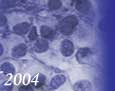
To perform quantification we used TEXCAN software (O. Ferrer, La Laguna, Tenerife, Spain), it allows the study of smears stained with progressive hematoxylin 5. Nuclei were visualized either through high resolution and a 60 x wet objective, nucleus by nucleus, or through a 40 x objective, which permits a greater number of cells in each view. We also used a black-and-white, charge-coupled device video camera for comparing shades of grey and a person-al computer.
A minimum of 100 study and 25 control cells were studied. The controls were stroma cells or lymphocytes and in all cases were internal controls, thus undergoing the same physical and chemical processes as did the study cells.
From each case studied we obtained a histogram of the study cell and of the control and compared ploidy in both. The histogram yielded the ploidy value, coefficient of variation and mean optical den-sity related to the modal peak of the G0-G1 and G2M phases. From those data we selected three cel-lular phases for study: G0-G1, S and G2M.
The software produces a prognosis index that gives values derived from quantification, such as the 5c exceeding rate and entropy value (E), or dispersion of the histogram.
The software offers other indexes and grades of malignancy, such as the morphometric prognostic index, malignancy index of Bócking and malig-nancy grade. They are based on histologic parame-ters, such as tumor size, mitotic activity index and existence of affected ganglioris. If our samples are compared to in vivo FNAB tumor characteristics, the size will change between palpation and the macroscopic tumor portion excised. The number of affected ganglions will differ. We did not use MAI.We did not consider the above factors in evaluating survival. Our evaluations were derived from the type of histogram.
|
|
|
|
|
|
|
Resultados
|
|
|
|
35 cases were collected and distributed in three groups as follows:
Group one: includes 6 cases with a first cytological diagnosis of dysplasia which fiberendoscopy and histology classified as cancer and latter confirmed by image analysis.
Group two: corresponds to cases considered as benign lesions by fiberendoscopy and biopsy but diagnosed as dyslasia by cytology and in which malignancy was ratified by image analysis. 14 cases are included in this group.
Group three: regards to dysplastic lesions by cytology but diagnosed as benign lesions by fiberendoscopy and biopsy as well as by image analysis. 15 cases belong to this group.
Histograms correspondent to group 3, since it is fully composed of benign cases, are all of them, type 0.
In the group 1, the image analysis shows a total agreement with pathology and fiberendoscopy, since the whole sample yields cancer type histograms, being in five cases (83,3%) types III and IV, representative of high grade of malignancy. All the patients were considered as having a malignant neoplasm and treated as that, obtaining the following survival rates:
Type I (low grade of malignnacy): 1case; currently alive without malignancy
Type III (high grade of malignancy): 2 cases; one patient died one year after diagnosis, the second patient was diagnosed and treated at 25-years of age, living free of disease at present.
Type IV (high grade of malignancy): 3 cases; only one patient is alive, the other two died one and five years after diagnosis.
These survival indices agrees with the data found for the well established tumors and with the references shown in the international literature.
For group three there is total concordance in between the four diagnostic techniques, finding no cancer cases. Survival for all patients varies from 11 to 19 years, with death caused by events different than cancer.
The group 2 shows for the type I histogram five cases, out of which 1 died due a mucinous adenocarcinoma of the colon, another one suffered surgical treatment and is currently alive with Crohn´s disease, the remaining behave as follows: 1 survived for 11 years, 1 survived for 14 years and died by different cause than cancer, the last one is currently alive.
For the type III (high grade) there are 8 cases, out of which 5 were exitus with survival rates among 1 and 5 years. Regarding the other 3, two suffered surgery and all of them are alive by year 2004.
Out of type IV (high grade) there is only one case who died due a gastric adenocarcinoma.
Summarazing, in this group 2, in which fiberendoscopy and biopsy failed to detect cancer, and cytology pointed out just dysplasia, 7 cases finally developed cancer and died (50% of the sample) being the malignant behaviour detected only by image cytometry. The other 7 cases in which image analysis indicated malignancy had better prognosis and high survival rates, since they were low grade of malignancy neoplasms.
Referring to the total 35 cases studied the amount of deaths imputables to cancer are 10, which means a 28,6%.
|
|
|
|
|
|
|
Discusión
|
|
|
|
Cytometry was developed more than 50 years ago, since then several hundreds of works have been published establishing the correlation between aneuploidy and cancer 1,2. In the 1980´s studies on gastric cancer began to appear 6,8, and few years later some investigators 9,10decided to focus on pre-malignant or dysplastic lessions rather than cancer. Their reports over different organs and lessions encouraged us to study benign entities of the female breast to demonstrate its bilogical behaviour 11. The present investigation aimed to assess the value of image cytometry in dysplasias of the gastrointestinal tract, and its possible role in evaluating the risk of malignant transformation of such lessions. In 1996 Marrero et al.10 concluded that cellular DNA content was abnormal at an early stage in dysplasia and stated that DNA analysis could be an important tool to select high-risk patients for screening. Their work presented a limitation since it was carried out in postgastrectomy patients; nevertheless in the same year other authors 12 published an interesting paper correlating endoscopy, histopathology and DNA flow cytometry, in which they indicated the need to biopsy and study the DNA in patients with gastric dyspepsia. The majority of authors 13,14 use flow cytometry to determine the DNA parameters of ploidy and S-phase-fraction, since our experience in assessing DNA by image cytometry in many cancerous lessions has been really possitive 3,4 we prefer this technique better than flow cytometry, else it allows more accurate histograms, eventhough the proccedure is slower. For this investigation we used image cytometry applied over cytological samples obtained through endoscopy, comparing the results with the histopathology of the biopsy specimen taken in the same act. There is very little published about image cytometry in the gastrointestinal tract precancerous lesions, two Chinese 15,16 authors refer in their findings that aneuploidy might be a marker of malignant change, very similar results have been found in the recent investigations about cytometry in gastrointestinal stromal tumours 17.When dealing with lesions located in the small and large bowel, the references are scarce and fail to obtain significant results 18.
In conclusion: this study proves that DNA quantification by image analysis in gastrointestinal tract dysplasias diagnosed by cytology is a very reliable tool in order to determine the real biological behaviour of the lesion, thus, allowing to be ahead a possible evolution to cancer. This technique also induces a more accurate differentiation between low and high grade of malignancy, and might be the unique prognostic factor that indicates such high-grade lesions, giving advice of more adequate therapeutic protocols, leading to closer follow-up or immediate surgical treatment if necessary. Supported by the results shown in this paper, we strongly recommend the practice of this diagnostic protocol –fiberendoscopy/cytology/biopsy/image analysis- to achieve a real knowledge of the biological behaviour of gastrointestinal lesions and allow a more adequate therapeutic guide.
|
|
|
|
|
|
|
Bibliografia
|
|
|
|
1. Auer G, Caspersson T, Wallgren A. DNA content and survival in mammary carcinoma. Anal Quant Cytol Histol 1980; 2(3): 161-166
2. Williams R A, Baak J P, Meijer G A, Charlton I G. Testing the value of a measurement protocol for DNA image cytometry and comparing different cytologic preparations. Anal Quant Cytol Histol 1996 Dec; 18(6): 429-37
3. Azua J, Romeo P, Morales M, Castiella T, Azua J: DNA quantification as a prognostic factor in gastric adenocarcinoma. Anal Quant Cytol Histol 1998 Jun; 20(3): 221-4
4. Azua J, Romeo P, Morales M, Castiella T, Azua J: DNA quantification as a prognostic factor in colorectal adenocarcinoma. Anal Quant Cytol Histol 1999 Dec; 21(6): 468-72
5. Schulte E K, Fink D K. Hematoxylin staining in quantitative DNA cytometry: an image analysis study. Anal Cell Pathol 1995 Dec; 9(4): 257-68
6. Ballatyne KC, James PD, Robin RA, Baldwin RW, Hardcastle JD. Flow cytomteric analysis of the DNA content of gastric cancer. Br J Cancer 1987;56:52-4
7. De Aretxablala X, Yonemura Y, Sugiyama K, Hirose N, Kumai T, Fushida S. DNA ploidy in gastric cancer and its relation to prognosis. Br J Cancer 1988;58:81-4
8. Filipe MI, Rosa J, Sandy A, Imrie PR, Ormerod MG, Morris RW. Is DNA ploidy and proliferative activity of prognostic value in advanced gastric carcinoma? Hum Pathol 1991;22:373-8
9. Macartney JC, Camplejohn RS. DNA flow cytometry of histological material from dysplastic lesions of human gastric mucosa. J Pathol 1986;150:113-8
10. Marrero JM, de Caestecker JS, Corbishley CM, McCormick C, Northfield TC. Gastric DNA content in postgastrectomy patients. Relationship to mucosal dysplasia. Cancer 1996;77:19-24
11. Azua Romeo J, de Azua Blanco J, Romeo P. Image cytometry in nonproliferative fibrocystic breast changes. Ploidy evaluation and epidemiologic study. Anal Quant Cytol Histol 2001 Apr; 23(2): 129-34
12. Abdel-Wahab M, Attallah AM, Elshal MF, Eldousoky I, Zalata KR, el-Gad el-Hak N, el-Ebidy G, Ezzat F. Correlation between endoscopy, histopathology, and DNA flow cytometry in patients with gastric dyspepsia. Hepatogastroenterology 1996;43:1313-20
13. Imada T, Yamamoto Y, Fukuzawa K, Rino Y, Suda T, Moriwaki Y, Akiyama H, Doi T, Kito F, Takemura H. Flow cytometric analysis of nuclear DNA heterogenity in gastric cancer. Jpn J Clin Oncol 1997;27:221-6
14. Chen BF, Marrogi AJ, Freeman SM, Clejan S. Gastric carcinoma: recent issues in prognostic factors. J La State Med Soc 1995;147:138-45
15. Shao L, Lei DN. Evaluation of DNA content in gastric dysplasia and carcinoma by image cytometry. Zhonghua Bing Li Xue Za Zhi 1989;18: 254-6
16. Zuo LF. DNA ploidy in gastric precancerous lesions. Zhonghua Zhong Liu Za Zhi 1991;13:180-3
17. Lerma E, Lee SJ, Tugues D, Oliva E, Gich I, Prat J. Ploidy of 36 stromal tumors of the gastrointestinal tract. A comparative study with flow cytometry and image analysis. 1994;16:435-40
18. Sasaki O, Soejima K. Fluorocytometric DNA analysis of borderline lesions in the large intestine. Gan To Kagaku Ryoho. 1993;20:717-20
|
|
|
|
|
|
|