|
|
Intracardiac unfrequent lessions.
Dr. Javier Azúa-Romeo
,
Dra Eva Moreno *
,
Dra Mayte Tovar Lázaro **
,
Dra. Isabel Marquina Ibáñez^
y
Dr. Jorge Alfaro Torres^
*Sº Cardiología. HUMS. Zaragoza
**MT. Tovar Lázaro. Sº Urgencias. MAZ. Zaragoza
^Sº Anatomía Patológica. HUMS. Zaragoza
España
|
|
|
|
|
|
|
|
Resumen
|
|
|
|
Hereby we present five pictures of rare cardiac tumours collected throughout the years; in addition we show an astonishment image of a massive cardiac involvement by a very unusual bacterial infection.
Primary tumours of the heart are really rare, with an incidence less than 0,3%; the most common one is cardiac myxoma, accounting for almost 50% of the benign tumours. Of the malignant tumours, angiosarcomas are the most frequent. Nevertheless, metastatic tumours are the most frequently encountered, with an incidence about 30-fold greater than primary neoplasms.
|
|
|
|
|
|
|
Discusión
|
|
|
|
It is difficult to assess the range of organisms involved in myocarditis, one study relates that viruses are the most usual organism responsible for infective myocarditis followed by bacteria (streptococo and staphilococo) and mycoplasma, resulting in over 99% of the collected cases.
Our cases correspond to biopsy or autopsy specimens, being most of them unsuspected findings in asymptomatic patients or studied by extra-cardiac-diseases.
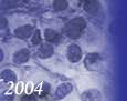
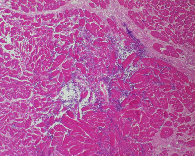
Case 1: elderly woman with nonspecific abdominal pain and orange muco-cutaneous discoloration. Patient died 12 hours after admittance. Image shows clusters of bacilli disrupting cardiac muscle fibres. Pathologic diagnosis: Clostridium tertium septicaemia.
Case 2: 50-years-old male, asymptomatic, with an encapsulated mass in the right ventricle. Microscopically was composed of mature fat cells with fibrous tissue and blood vessels. Pathologic diagnosis: cardiac lipoma.
Case 3: 36-year-old female with symptoms of pulmonary valve disease. Picture shows a papillary tumour that arose from the endocardium of the right ventricle. Pathologic diagnosis: papillary fibroelastoma.
Case 4: 20-year-old female that presented with irregular cardiomegaly and arrhythmia. Microscopic studies revealed a pleomorphic lesion with marked anaplasia with scattered rhabdomyoblasts. Pathologic diagnosis: rhabdomyosarcoma.
Case 5: elderly man with respiratory symptoms and congestive failure. Cardiac biopsy was performed showing nests of big, atypical, rounded cells with an inflammatory background. Pathologic diagnosis: metastatic carcinoma of the lung.
Case 6: Rhythm disturbances in 45-year-old male, image techniques demonstrated a diffuse cardiac neoplasm. Microscopically displayed a malignant epithelial pattern, lacking specific features. Immunohistochemical staining procedure favoured the diagnosis of liver carcinoma versus melanoma. CT-scan found a mass in the right lobe of the liver. Pathologic diagnosis: metastatic carcinoma of the liver.
We encourage the myocardial biopsy to achieve more accurate diagnosis.
|
|
|
|
|
|
|
Bibliografia
|
|
|
|
1. J. Azúa-Romeo, E. Moreno, J.P. Gomollón. Right atrial lithomixoma with extramedullary hematopoiesis. Heart2002; 88:10-10
2. JM Grasa, J Azúa-Romeo, MJ Borruel, A Milagro, MI Fuertes, JC Garcia-Zueco. Sepsis anaerobia fulminante en paciente diabética con fístula colecistoentérica asintomática. Revista Clínica Española 2002; 202:619-621
|
|
|
|
|
|
|
NOTA:
Esto es un foro médico profesional, que no tiene como objetivo ofrecer consejo médico o de salud. Los mensajes enviados a este foro solicitando estos consejos, no serán atendidos. La información científica ofrecida está refrendada por las referencias y bibliografía correspondientes y de su veracidad son responsables sus autores. La participación en este Congreso es gratuita.
Todos los derechos reservados (C) UNINET 2003
Versión 1.5
Si experimenta cualquier problema en la edición de su trabajo envie un mensaje a la siguiente dirección: info.conganat@infomed.sld.cu


 Zoom
Zoom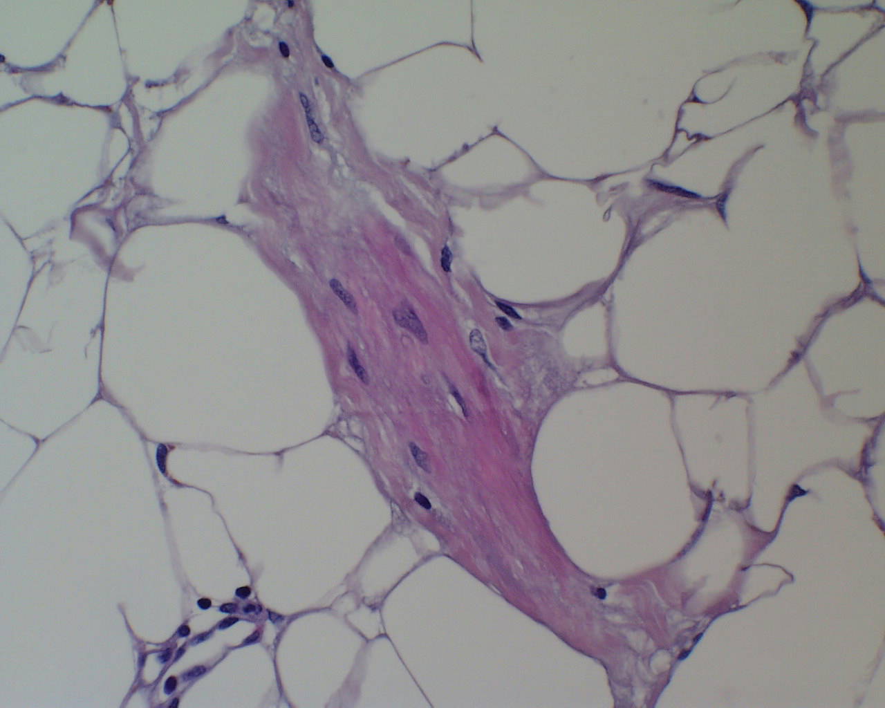 Zoom
Zoom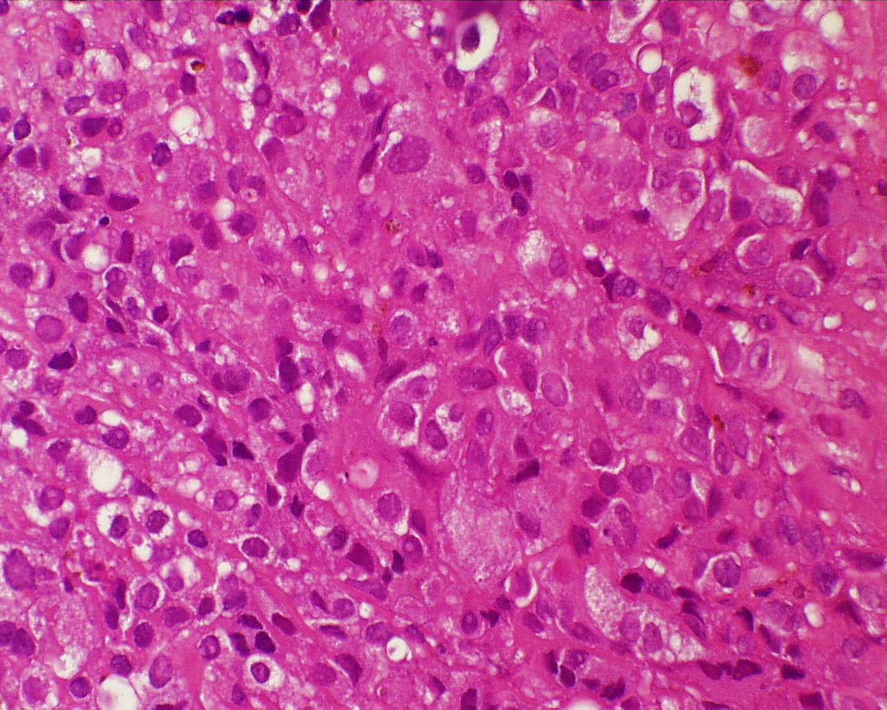 Zoom
Zoom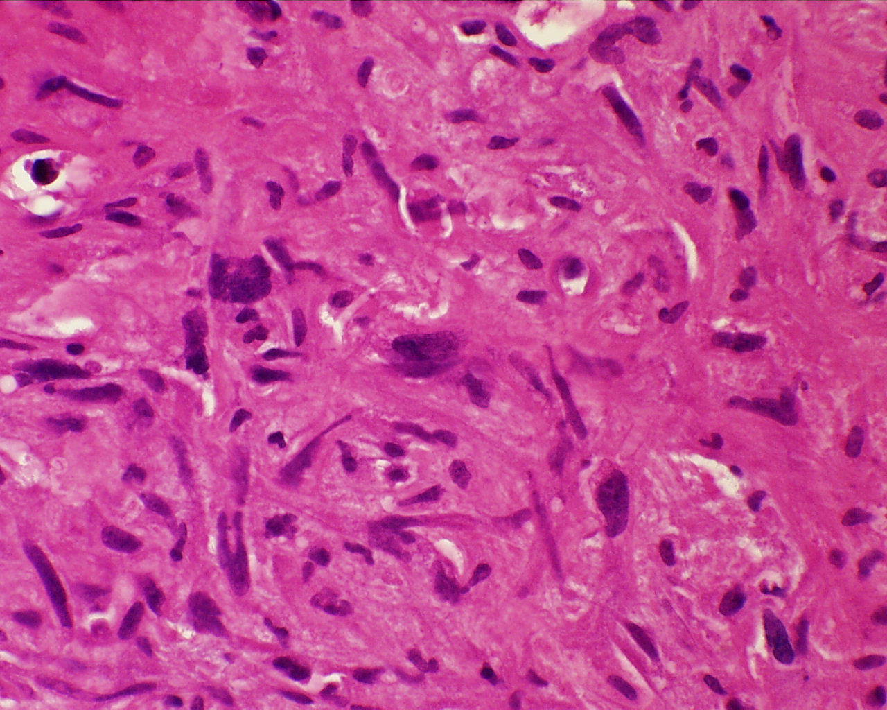 Zoom
Zoom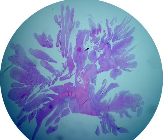 Zoom
Zoom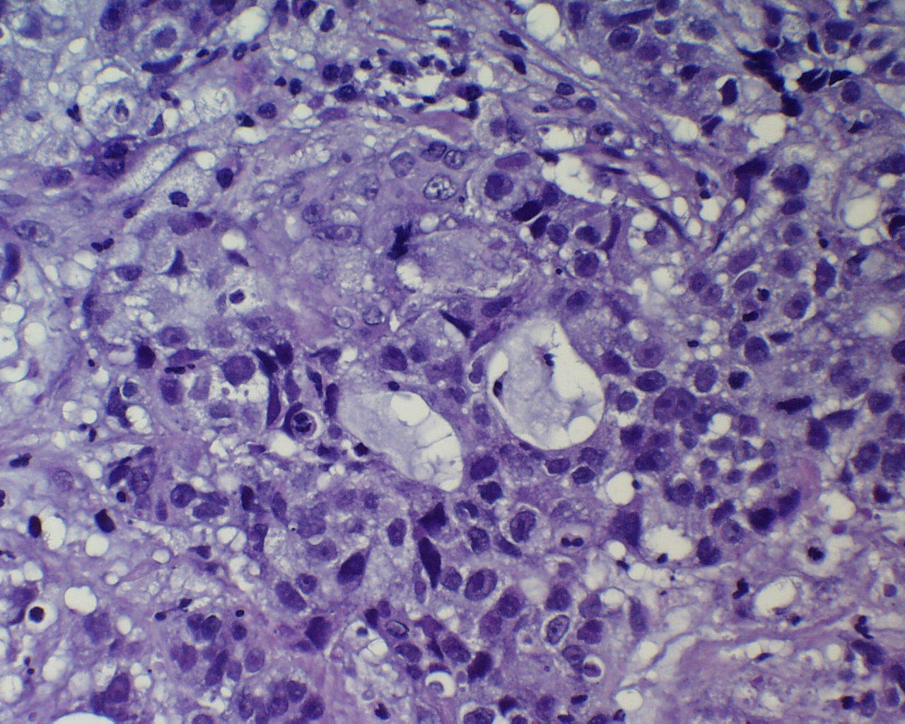 Zoom
Zoom